Carlson E J, Peterson E M, de la Maza L M
Department of Pathology, University of California, Irvine 92717.
Infect Immun. 1989 Feb;57(2):487-94. doi: 10.1128/iai.57.2.487-494.1989.
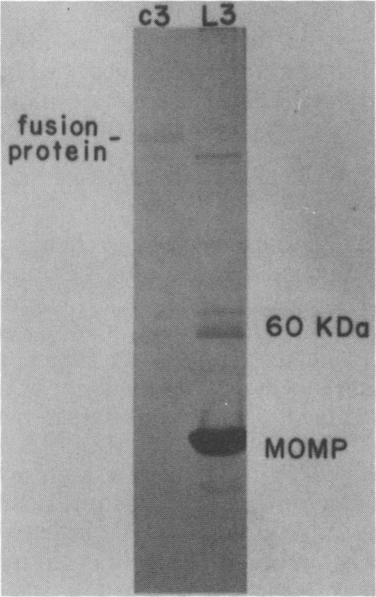
Chlamydia trachomatis L3 DNA was cloned and expressed in lambda gt11. A recombinant plaque that expressed an antigen that reacted with rabbit polyclonal antichlamydial L3 serum and with two monoclonal antibodies specific for serovars L3 and I was selected from this Chlamydia genomic library. The beta-galactosidase Chlamydia fusion protein was purified by immunoaffinity chromatography and injected into mice to produce monoclonal antibodies. These monoclonal antibodies reacted by Western (immuno-) blot with both the fusion protein and the major outer membrane protein from purified L3 elementary bodies. The chlamydial DNA fragment was shown by DNA sequence analysis to be 168 base pairs in length and to correspond to the constant regions 1 and 2 and the variable segment 1 of the major outer membrane protein gene. The recombinant chlamydial DNA fragment hybridized under stringent conditions by Southern and dot blot analysis exclusively with the DNA from the C- and C-related-complex C. trachomatis serovars.
沙眼衣原体L3 DNA被克隆并在λgt11中表达。从该衣原体基因组文库中筛选出一个重组噬菌斑,其表达的抗原能与兔抗衣原体L3多克隆血清以及两种分别针对血清型L3和I的单克隆抗体发生反应。通过免疫亲和层析法纯化β-半乳糖苷酶-衣原体融合蛋白,并将其注射到小鼠体内以产生单克隆抗体。这些单克隆抗体通过蛋白质免疫印迹法与融合蛋白以及纯化的L3原体的主要外膜蛋白发生反应。通过DNA序列分析表明,衣原体DNA片段长度为168个碱基对,对应于主要外膜蛋白基因的恒定区1和2以及可变区1。通过Southern印迹和斑点印迹分析,重组衣原体DNA片段在严格条件下仅与沙眼衣原体血清型C及其相关复合群的DNA杂交。