Markowski Jarosław, Jagosz-Kandziora Estera, Likus Wirginia, Pająk Jacek, Mrukwa-Kominek Ewa, Paluch Jarosław, Dziubdziela Włodzimierz
Department of ENT, Medical University of Silesia, Katowice, Poland.
Department of Human Anatomy, Medical University of Silesia, Katowice, Poland.
Med Sci Monit. 2014 Jun 16;20:988-94. doi: 10.12659/MSM.890433.
The aim of this study was to investigate the distribution of different types of primary orbital tumors, histopathological diagnosis, and postoperative complications.
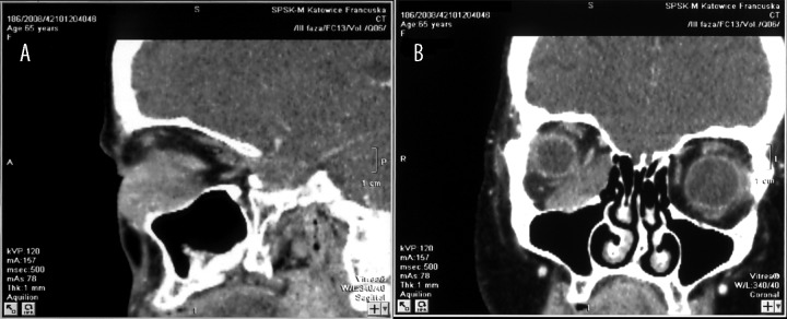
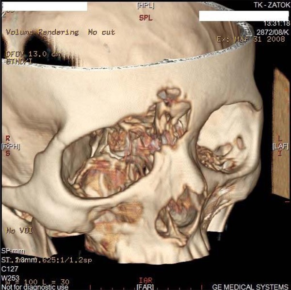
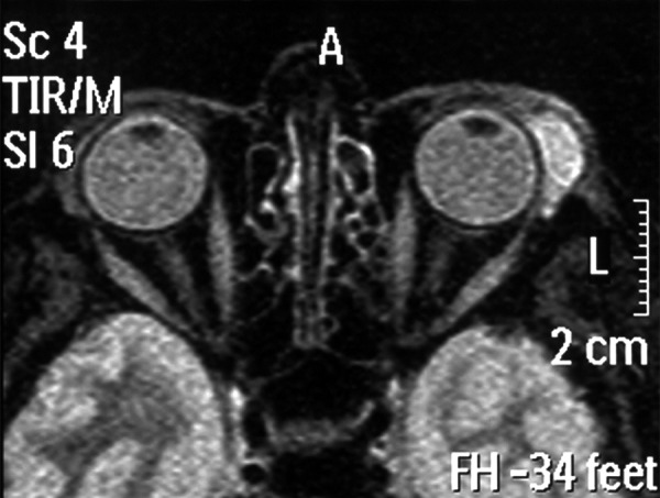
We analyzed 122 patients (68 women and 54 men) with orbital tumors, hospitalized in the ENT Department of the Medical University of Silesia in Katowice during 1990-2013. The patients were characterized in terms of anatomic, topographical, histopathological, and clinical parameters. The role of diagnostic imagining such as CT, NMR, and fine-needle aspiration (FNB) in preoperative diagnostics is discussed.
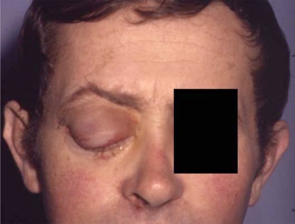
of FNB, cytological, and histopathological examination of the postoperative specimens were compared. Results There were 56 (46%) patients with malignant tumors, 42 (34%) with benign tumors, 19 (16%) with inflammatory tumors, and 5 patients (4%) had other tumors. In cases of malignant tumors, local recurrence up to 5 years was found in 36 (64.3%) cases. In the other 20 (35.7%) cases of malignant tumors, the patients remained under close follow-up in the outpatient clinic, without signs of local recurrence (follow-up 1-17 years). According to histopathological examination, malignant tumors were detected in 45.9% of patients and non-malignant tumor in 34.4% of patients. In 19.7% of patients, inflammatory and other types of tumors were diagnosed.
We characterized the occurrence and pathological profiles of orbital tumors. The tumor location, histopathological diagnosis, and postoperative complications give us important information for the diagnosis of tumor prior to biopsy or tumor resection and for the determination of the treatment strategy and possible complications after surgery.
本研究旨在调查不同类型原发性眼眶肿瘤的分布、组织病理学诊断及术后并发症。
我们分析了1990年至2013年期间在卡托维兹西里西亚医科大学耳鼻喉科住院的122例眼眶肿瘤患者(68例女性和54例男性)。对患者的解剖学、局部解剖学、组织病理学和临床参数进行了分析。讨论了CT、核磁共振成像(NMR)和细针穿刺活检(FNB)等诊断性影像学检查在术前诊断中的作用。
比较了FNB、细胞学检查及术后标本的组织病理学检查结果。结果显示,56例(46%)为恶性肿瘤患者,42例(34%)为良性肿瘤患者,19例(16%)为炎性肿瘤患者,5例(4%)为其他肿瘤患者。在恶性肿瘤病例中,36例(64.3%)在5年内出现局部复发。另外20例(35.7%)恶性肿瘤患者在门诊密切随访,无局部复发迹象(随访1至17年)。根据组织病理学检查,45.9%的患者检测出恶性肿瘤,34.4%的患者检测出非恶性肿瘤。19.7%的患者被诊断为炎性肿瘤和其他类型肿瘤。
我们对眼眶肿瘤的发生情况和病理特征进行了描述。肿瘤位置、组织病理学诊断及术后并发症为活检或肿瘤切除术前的肿瘤诊断、治疗策略的确定以及术后可能出现的并发症提供了重要信息。