Department of Mechanics and Engineering Science, College of Engineering, Peking University, Beijing 100871, China.
Head Face Med. 2014 Jun 19;10:24. doi: 10.1186/1746-160X-10-24.
Excessive compressive and shear stresses are likely related to condylar resorption and disc perforation. Few studies have reported the disc displacement and deformation during jaw opening. The aim of this study was to analyze stress distribution in a normal articular disc during the jaw opening movement.
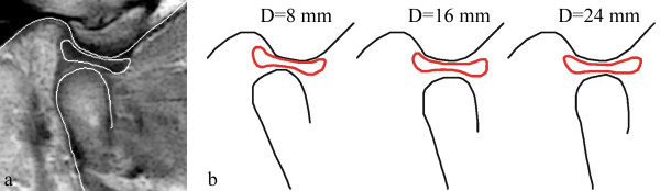
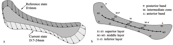
Bilateral MRI images were obtained from the temporomandibular joint of a healthy subject for the jaw opening displacement from 6 to 24 mm with 1 mm increments. The disc contour for the jaw opening at 6 mm was defined as the reference state, and was used to establish a two dimensional finite element model of the disc. The contours of the disc at other degrees of jaw opening were used as the displacement loading. Hyperelastic material models were applied to the anterior, intermediate and posterior parts of the disc. Stress and strain trajectories were calculated to characterize the stress/strain patterns in the disc.
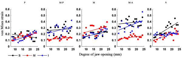
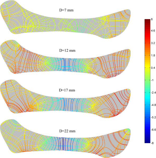
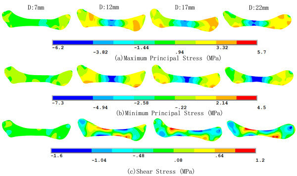
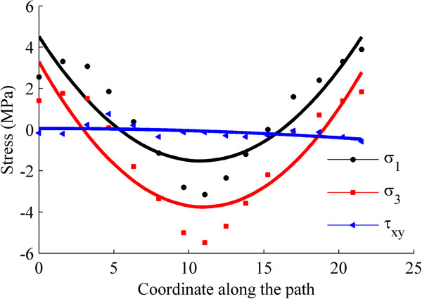
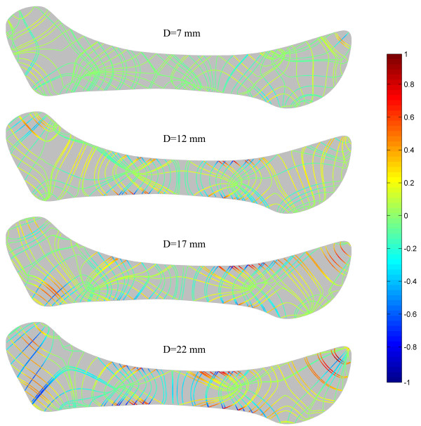
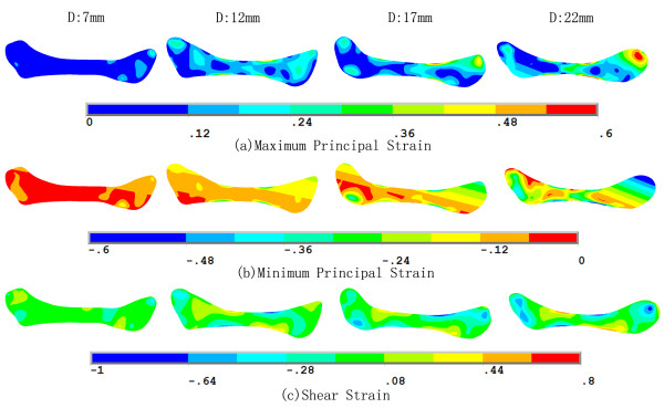
Both the maximum and minimum principal stresses were negative in the intermediate zone, therefore, the intermediate zone withstood mainly compressive stress. On the contrary, the maximum and minimum principal stresses were most positive in the anterior and posterior zones, which meant that the anterior and posterior bands suffered higher tensile stresses. The different patterns of stress trajectories between the intermediate zone and the anterior and posterior bands might be attributed to the effect of fiber orientation. The compression of the intermediate zone and stretching of the anterior and posterior bands caused high shear deformation in the transition region, especially at the disc surfaces.
The stress and strain remained at a reasonable level during jaw opening, indicating that the disc experiences no injury during functional opening movements in a healthy temporomandibular joint.
过大的压缩和剪切应力可能与髁突吸收和盘穿孔有关。很少有研究报道过在张口过程中盘的位移和变形。本研究旨在分析正常关节盘中的应力分布在张口运动。
从健康受试者的颞下颌关节获得双侧 MRI 图像,用于张口从 6 毫米到 24 毫米,以 1 毫米为增量。6 毫米时的盘轮廓被定义为参考状态,并用于建立盘的二维有限元模型。在其他张口程度下的盘轮廓被用作位移加载。超弹性材料模型应用于盘的前、中和后区。计算应力和应变轨迹,以表征盘内的应力/应变模式。
中间区的最大和最小主应力均为负,因此中间区主要承受压缩应力。相反,前区和后区的最大和最小主应力最为正值,这意味着前区和后区带承受较高的拉伸应力。中间区与前区和后区带之间的不同的应力轨迹模式可能归因于纤维方向的影响。中间区的压缩和前区和后区带的拉伸导致过渡区的高剪切变形,特别是在盘表面。
在张口过程中,应力和应变保持在合理的水平,表明在健康的颞下颌关节的功能张口运动中,盘不会受到损伤。