Takeuchi Kayo, Ono Ayako, Yamada Atsushi, Toyooka Mariko, Takahashi Takahiro, Shigematsu Yoshiki, Ohta Makoto, Sagoh Tadashi
Department of Radiology, Fukui Red Cross Hospital, Fukui, Japan.
Department of Chest Surgery, Fukui Red Cross Hospital, Fukui, Japan.
Pol J Radiol. 2014 Jun 12;79:145-9. doi: 10.12659/PJR.890662. eCollection 2014.
This case report describes two cases of extralobar pulmonary sequestration in adults with and without torsion/necrosis.
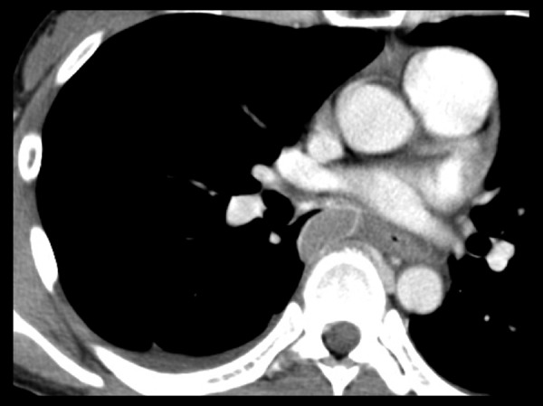
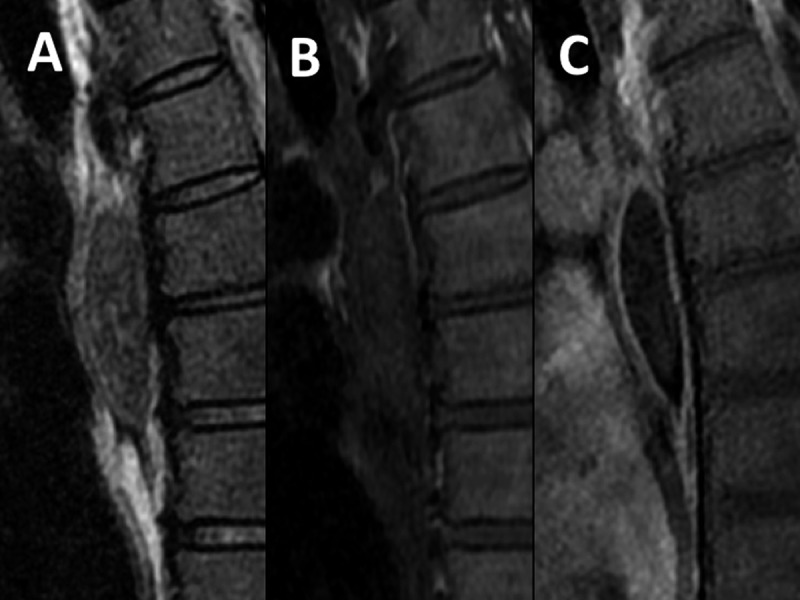
Non-complicated extralobar pulmonary sequestration was found incidentally in a 50-year-old asymptomatic woman (Case 1), diagnosed with the presence of a branching structure in a mass lesion and blood supply from the right inferior phrenic artery. Another case of a 38-year-old woman presented with a sudden onset of back pain caused by extralobar pulmonary sequestration with torsion/necrosis (Case 2). A 4-cm fusiform mass in the paravertebral region showed enhancement in the peripheral rim only, and no feeding artery. These were the same as it had been reported typical findings in extralobar pulmonary sequestration with necrosis. On magnetic resonance imaging, the masses in both cases showed inhomogeneous low signal and branching high signal on T2-weighted images. That was characteristic for a stroma without dilated alveoli as a solid part and dilated alveoli as fluid regions.
By comparing those two cases, we came to a conclusion that only T2-weighted imaging reflects the native structure, even after infarction. Although differentiation from a cystic tumor with hemorrhage or infection can be problematic, inhomogeneous low signal and branching high signal on T2-weighted images may help us distinguish extralobar pulmonary sequestration from other cystic lesions.
本病例报告描述了两例成人肺叶外型肺隔离症,其中一例伴有扭转/坏死,另一例未伴有扭转/坏死。
一名50岁无症状女性偶然发现非复杂性肺叶外型肺隔离症(病例1),在肿块病变中诊断出存在分支结构且血供来自右下膈动脉。另一例38岁女性因肺叶外型肺隔离症伴扭转/坏死突然出现背痛(病例2)。椎旁区域一个4厘米的梭形肿块仅在周边边缘有强化,且无供血动脉。这些与已报道的肺叶外型肺隔离症伴坏死的典型表现相同。在磁共振成像上,两例病例的肿块在T2加权图像上均显示不均匀低信号和分支状高信号。这是无扩张肺泡的间质作为实性部分、扩张肺泡作为液体区域的特征表现。
通过比较这两例病例,我们得出结论,即使在梗死之后,只有T2加权成像能反映其原生结构。尽管与伴有出血或感染的囊性肿瘤进行鉴别可能存在问题,但T2加权图像上的不均匀低信号和分支状高信号可能有助于我们将肺叶外型肺隔离症与其他囊性病变区分开来。