Chou Ming-Li, Burnouf Thierry, Wang Tsung-Jen
Graduate Institute of Medical Sciences, College of Medicine, Taipei Medical University, Taipei, Taiwan, R.O.C.
Graduate Institute of Biomedical Materials and Tissue Engineering, Taipei Medical University, Taipei, Taiwan, R.O.C.
PLoS One. 2014 Jun 19;9(6):e99145. doi: 10.1371/journal.pone.0099145. eCollection 2014.
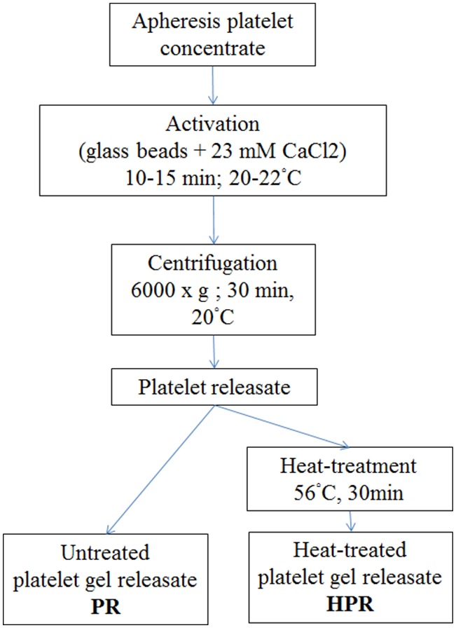
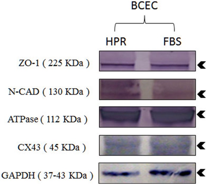
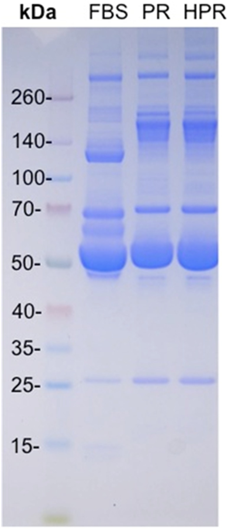
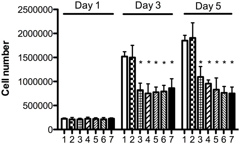
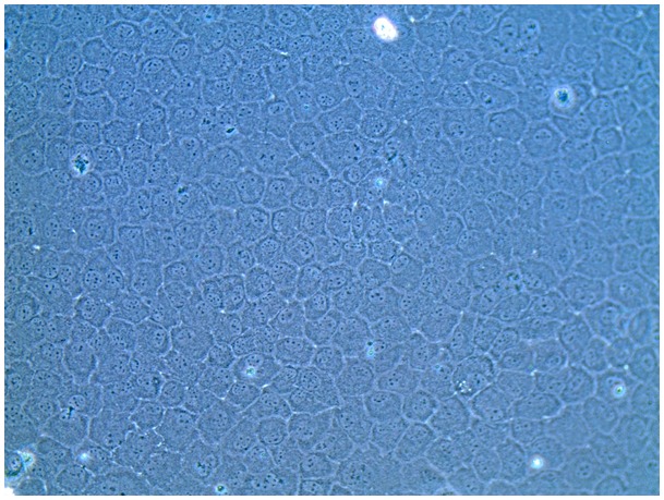
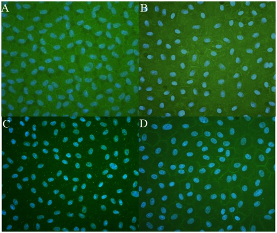
Clinical-grade ex vivo expansion of corneal endothelial cells can increase the availability of corneal tissues for transplantation and treatment of corneal blindness. However, these cells have very limited proliferative capacity. Successful propagation has required so far to use very complex growth media supplemented with fetal bovine serum and other xenocomponents. We hypothesized that human platelet releasates rich in multiple growth factors, and in particular neurotrophins, could potentially be a useful supplement for ex vivo expansion of corneal endothelium cells due to their neural crest origin. Platelet releasates were prepared by calcium salt activation of apheresis platelet concentrates, subjected or not to complement inactivation by heat treatment at 56°C for 30 minutes. Platelet releasates were characterized for their content in proteins and were found to contain high amount of growth factors including platelet-derived growth factor-AB (30.56 to 39.08 ng/ml) and brain-derived neurotrophic factor (30.57 to 37.11 ng/ml) neurotrophins. We compared the growth and viability of corneal endothelium cells in DMEM-F12 medium supplemented with different combinations of components, including 2.5%∼10% of the platelet releasates. Corneal endothelium cells expanded in platelet releasates exhibited good adhesion and a typical hexagonal morphology. Their growth and viability were enhanced when using the complement-inactivated platelet releasate at a concentration of 10%. Immunostaining and Western blots showed that CECs maintained the expressions of four important membrane markers: Na-K ATPase α1, zona occludens-1, phospho-connexin 43 and N-cadherin. In conclusion, our study provides the first proof-of-concept that human platelet releasates can be used for ex vivo expansion of corneal endothelium cells. These findings open a new paradigm for ex vivo propagation protocols of corneal endothelium cells in compliance with good tissue culture practices and regulatory recommendations to limit the use of xenogenic materials.
临床级角膜内皮细胞的体外扩增可增加用于角膜移植和治疗角膜盲的角膜组织的可用性。然而,这些细胞的增殖能力非常有限。迄今为止,成功的增殖需要使用非常复杂的生长培养基,并添加胎牛血清和其他异种成分。我们推测,富含多种生长因子,特别是神经营养因子的人血小板释放物,由于其神经嵴起源,可能是角膜内皮细胞体外扩增的有用补充物。通过对单采血小板浓缩物进行钙盐激活制备血小板释放物,56℃热处理30分钟进行或不进行补体灭活。对血小板释放物的蛋白质含量进行了表征,发现其含有大量生长因子,包括血小板衍生生长因子-AB(30.56至39.08 ng/ml)和脑源性神经营养因子(30.57至37.11 ng/ml)等神经营养因子。我们比较了在添加不同成分组合(包括2.5%至10%的血小板释放物)的DMEM-F12培养基中角膜内皮细胞的生长和活力。在血小板释放物中扩增的角膜内皮细胞表现出良好的粘附性和典型的六边形形态。当使用浓度为10%的补体灭活血小板释放物时,它们的生长和活力得到增强。免疫染色和蛋白质印迹显示,角膜内皮细胞维持四种重要膜标志物的表达:钠钾ATP酶α1、紧密连接蛋白-1、磷酸化连接蛋白43和N-钙粘蛋白。总之,我们的研究首次提供了概念验证,即人血小板释放物可用于角膜内皮细胞的体外扩增。这些发现为角膜内皮细胞的体外增殖方案开辟了新的范例,符合良好的组织培养规范和限制使用异种材料的监管建议。