Jayi Sofia, Laadioui Meriem, El Fatemi Hind, Fdili Fatima Zohra, Bouguern Hakima, Chaara Hikmat, Laamarti Afaf, Melhouf My Abdelilah
Department of Gynecology and Obstetrics, University Hospital of Fez, Sidi Mohammed Ben Abdellah University, 37-39, Lotissement Asmae, Route Ain Chqef, Fez, Morocco.
J Med Case Rep. 2014 Jun 18;8:203. doi: 10.1186/1752-1947-8-203.
Vulvar lipoma is a rare tumor localization and only a few cases have been reported. The clinical characteristics of vulvar lipoma are well known. However, it is important to distinguish lipomas from liposarcomas. We report a case of vulvar lipoma and discuss its clinical features, including diagnostic aspects, with emphasis on histopathological evaluation of all excised lesions. We also report and discuss patient management and treatment outcomes.
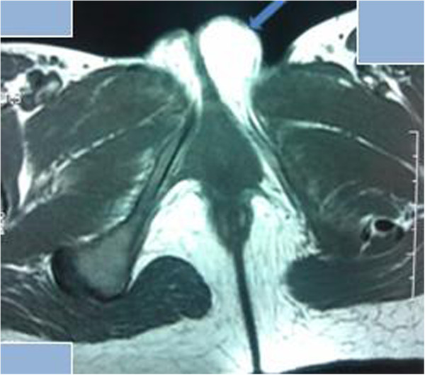
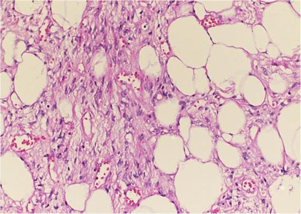
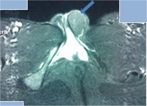
We report the case of a 27-year-old Moroccan woman. Our patient presented with a painless and slow-growing right vulvar mass that had evolved over one year, which had suddenly become uncomfortable when walking. A physical examination revealed a single soft and pasty mass in her left labium majus, which could be mobilized under her skin towards her mons pubis. The largest dimension of the mass measured 6cm. Magnetic resonance imaging showed a homogenous hyperintense mass with a well-defined contour in her left labium majus; a fat-suppressed magnetic resonance image demonstrated a marked signal intensity decrease. The mass was completely removed surgically. A histological examination revealed a circumscribed benign tumor composed of mature adipocytes, confirming the diagnosis of vulvar lipoma.
Vulvar lipomas must be differentiated from liposarcomas, which demonstrate very similar clinical and imaging profiles. The final diagnosis should be based on histopathological evaluation. A precise diagnosis should allow for appropriate surgical treatment.
外阴脂肪瘤是一种罕见的肿瘤部位,仅有少数病例报道。外阴脂肪瘤的临床特征已为人熟知。然而,将脂肪瘤与脂肪肉瘤区分开来很重要。我们报告一例外阴脂肪瘤病例,并讨论其临床特征,包括诊断方面,重点是对所有切除病变的组织病理学评估。我们还报告并讨论了患者管理和治疗结果。
我们报告一例27岁摩洛哥女性病例。我们的患者表现为右侧外阴无痛性、生长缓慢的肿块,已发展一年,行走时突然变得不适。体格检查发现左侧大阴唇有一个单一的柔软、糊状肿块,可在皮下向耻骨联合移动。肿块最大直径为6厘米。磁共振成像显示左侧大阴唇有一个轮廓清晰的均匀高信号肿块;脂肪抑制磁共振图像显示信号强度明显降低。该肿块通过手术完全切除。组织学检查显示为一个由成熟脂肪细胞组成的边界清楚的良性肿瘤,证实为外阴脂肪瘤。
外阴脂肪瘤必须与脂肪肉瘤相鉴别,后者表现出非常相似的临床和影像学特征。最终诊断应基于组织病理学评估。准确的诊断应有助于进行适当的手术治疗。