Guber Josef, Josifova Tatjana, Henrich Paul Bernhard, Guber Ivo
Sutton Eye Hospital, Epsom and St Helier University Hospitals, London, UK.
Department of Ophthalmology, University of Basel, Basel, Switzerland.
Open Ophthalmol J. 2014 May 16;8:3-6. doi: 10.2174/1874364101408010003. eCollection 2014.
To identify OCT-based anatomical features and clinical characteristics for poor central retinal thickness (CRT) response to ranibizumab in neovascular age-related macular degeneration (AMD).
Investigating our electronic patient records (Eyeswide), patients with neovascular AMD treated with intravitreal injections of 0.5mg/0.05ml ranibizumab were identified and their notes reviewed. Data collected included gender, age, initial best-corrected visual acuity (BCVA), prior photodynamic therapy, lesion type (classic versus occult), type of macular edema (intraretinal fluid, subretinal fluid, pigment epithelium detachment) and the total number of previous ranibizumab injections.
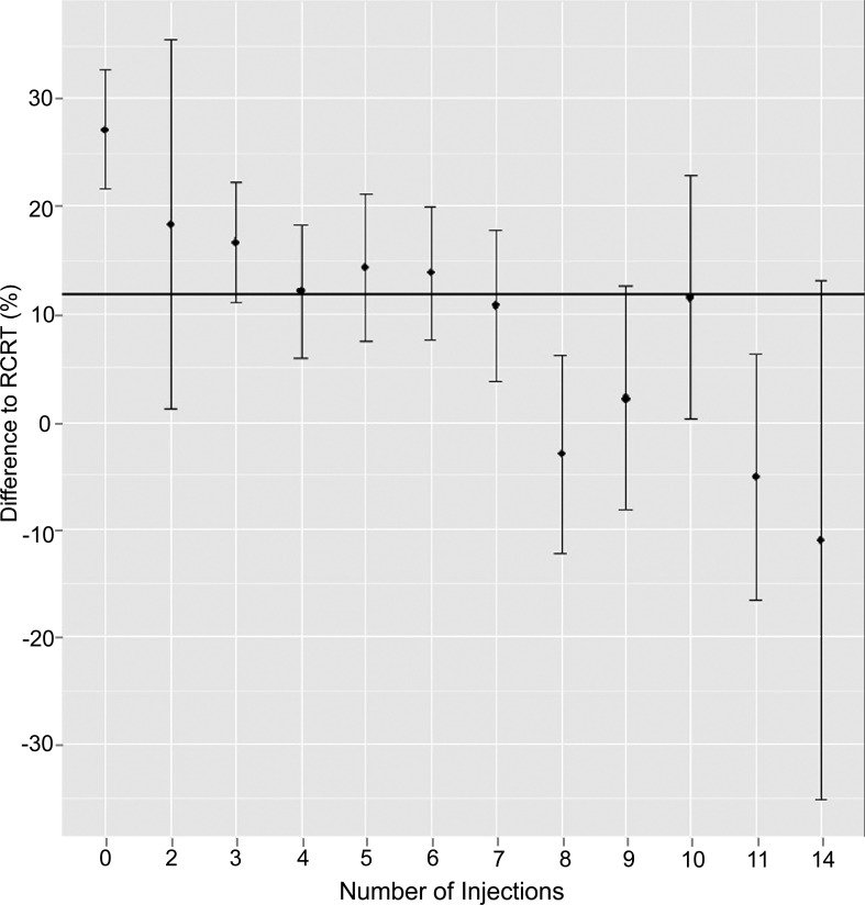
A total of 210 eyes of 182 patients with neovascular AMD were identified. Mean follow-up time was 1.34 years (SD ± 0.77). Central retinal thickness reduction in women was significantly inferior to that in men (p=0.05). Patients with cystoid type macular edema had significantly greater reduction in CRT compared to patients with subretinal fluid (p<0.001) or pigment epithelium detachment (p<0.001). The percentage drop of CRT was no longer statistically significant after the sixth injection. Age, initial BCVA, prior photodynamic therapy and lesion type had no statistically effect on CRT response.
Risk factors for poor central retinal thickness response to ranibizumab include female gender and patients with predominant subretinal fluid or pigment epithelium detachment. Furthermore, the anatomical response decreased after the sixth injection of ranibizumab.
确定基于光学相干断层扫描(OCT)的解剖学特征及临床特征,以了解新生血管性年龄相关性黄斑变性(AMD)患者对雷珠单抗治疗后中心视网膜厚度(CRT)反应不佳的情况。
通过查阅电子病历(Eyeswide),确定接受玻璃体内注射0.5mg/0.05ml雷珠单抗治疗的新生血管性AMD患者,并对其病历进行回顾。收集的数据包括性别、年龄、初始最佳矫正视力(BCVA)、既往光动力疗法、病变类型(典型与隐匿性)、黄斑水肿类型(视网膜内液、视网膜下液、色素上皮脱离)以及既往雷珠单抗注射的总数。
共确定了182例新生血管性AMD患者的210只眼。平均随访时间为1.34年(标准差±0.77)。女性患者的中心视网膜厚度降低明显低于男性(p=0.05)。与视网膜下液(p<0.001)或色素上皮脱离(p<0.001)的患者相比,囊样黄斑水肿患者的CRT降低明显更大。第六次注射后,CRT的下降百分比不再具有统计学意义。年龄、初始BCVA、既往光动力疗法和病变类型对CRT反应无统计学影响。
对雷珠单抗治疗后中心视网膜厚度反应不佳的危险因素包括女性以及以视网膜下液或色素上皮脱离为主的患者。此外,雷珠单抗第六次注射后解剖学反应降低。