• 文献检索
  • 文档翻译
  • 深度研究
  • 学术资讯
  • Suppr Zotero 插件Zotero 插件
  • 邀请有礼
  • 套餐&价格
  • 历史记录
应用&插件
Suppr Zotero 插件Zotero 插件浏览器插件Mac 客户端Windows 客户端微信小程序
定价
高级版会员购买积分包购买API积分包
服务
文献检索文档翻译深度研究API 文档MCP 服务
关于我们
关于 Suppr公司介绍联系我们用户协议隐私条款
关注我们

Suppr 超能文献

核心技术专利:CN118964589B侵权必究
粤ICP备2023148730 号-1Suppr @ 2026

文献检索

告别复杂PubMed语法,用中文像聊天一样搜索,搜遍4000万医学文献。AI智能推荐,让科研检索更轻松。

立即免费搜索

文件翻译

保留排版,准确专业,支持PDF/Word/PPT等文件格式,支持 12+语言互译。

免费翻译文档

深度研究

AI帮你快速写综述,25分钟生成高质量综述,智能提取关键信息,辅助科研写作。

立即免费体验

门静脉系统及其属支的径向超声内镜评估。

Portal venous system and its tributaries: a radial endosonographic assessment.

机构信息

Department of Gastroenterology, Jaswant Rai Specialty Hospital, Saket, Meerut, India.

Department of Anatomy, L.L.R.M., Medical College, Meerut 250001, U.P., India.

出版信息

Endosc Ultrasound. 2012 Jul;1(2):96-107. doi: 10.7178/eus.02.008.

DOI:10.7178/eus.02.008
PMID:24949345
原文链接:https://pmc.ncbi.nlm.nih.gov/articles/PMC4062210/
Abstract

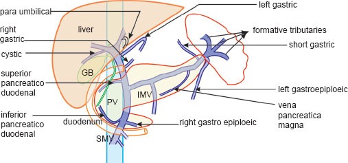
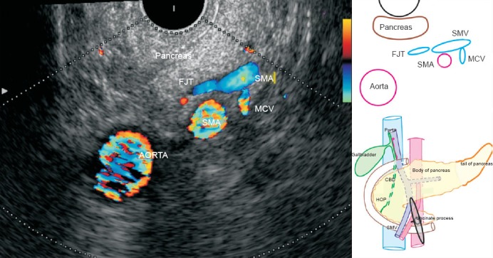
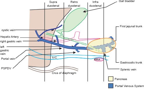
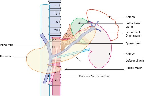
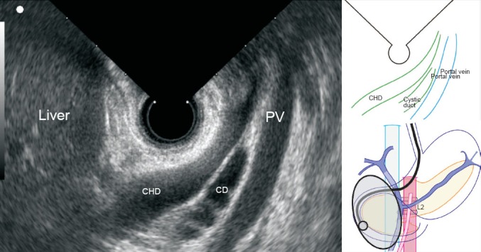
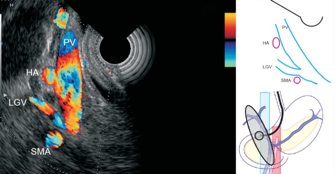
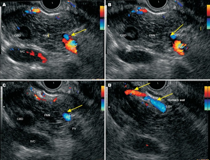
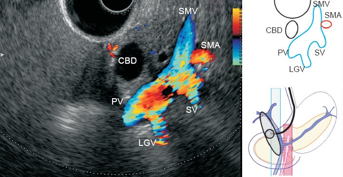
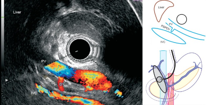
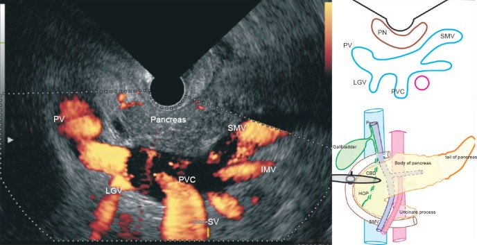
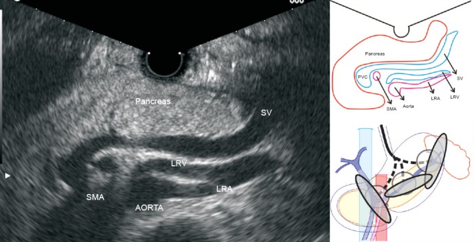
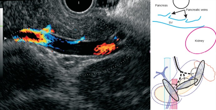
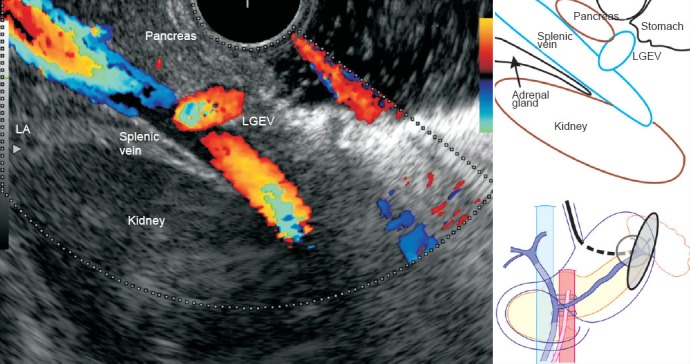
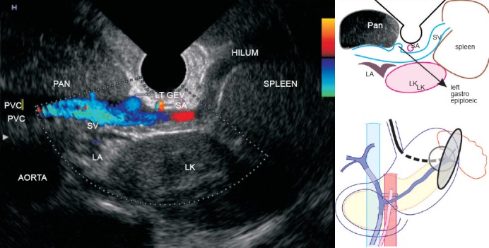
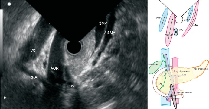
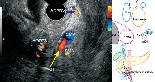
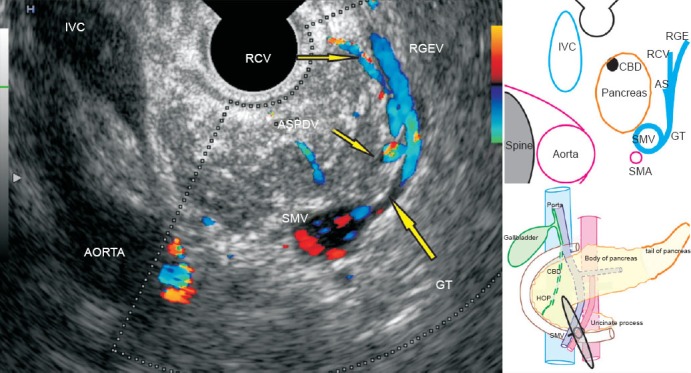
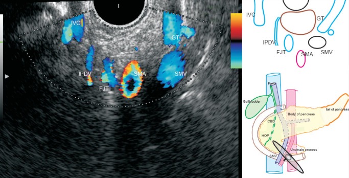
The use of Color Doppler in endosonography has enabled detailed real-time assessment of the abdominal vasculature. Standard stations are used during the routine evaluation on endosonography. However, the imaging techniques do not describe the vascular imaging of the portal venous system and its tributaries, in detail. This article demonstrates the normal findings on the portal venous system and its tributaries using radial endosonography.

摘要

彩色多谱勒在超声内镜中的应用使得对腹部脉管系统的详细实时评估成为可能。在常规的超声内镜评估中使用标准的检查部位。然而,这些成像技术并不能详细描述门静脉系统及其属支的血管成像。本文使用径向超声内镜显示门静脉系统及其属支的正常表现。

https://cdn.ncbi.nlm.nih.gov/pmc/blobs/9cb3/4062210/d1146c0922fd/EUS-1-96-g022.jpg
https://cdn.ncbi.nlm.nih.gov/pmc/blobs/9cb3/4062210/89f0c8900fd6/EUS-1-96-g001.jpg
https://cdn.ncbi.nlm.nih.gov/pmc/blobs/9cb3/4062210/88262c1278fb/EUS-1-96-g002.jpg
https://cdn.ncbi.nlm.nih.gov/pmc/blobs/9cb3/4062210/ce4e229e5a67/EUS-1-96-g003.jpg
https://cdn.ncbi.nlm.nih.gov/pmc/blobs/9cb3/4062210/8d071abdcdc9/EUS-1-96-g004.jpg
https://cdn.ncbi.nlm.nih.gov/pmc/blobs/9cb3/4062210/56e713127693/EUS-1-96-g005.jpg
https://cdn.ncbi.nlm.nih.gov/pmc/blobs/9cb3/4062210/be548e317ea8/EUS-1-96-g006.jpg
https://cdn.ncbi.nlm.nih.gov/pmc/blobs/9cb3/4062210/7e610bfe98e1/EUS-1-96-g007.jpg
https://cdn.ncbi.nlm.nih.gov/pmc/blobs/9cb3/4062210/3bd1aab51460/EUS-1-96-g008.jpg
https://cdn.ncbi.nlm.nih.gov/pmc/blobs/9cb3/4062210/9a886fcbe350/EUS-1-96-g009.jpg
https://cdn.ncbi.nlm.nih.gov/pmc/blobs/9cb3/4062210/c2528bba79bd/EUS-1-96-g010.jpg
https://cdn.ncbi.nlm.nih.gov/pmc/blobs/9cb3/4062210/803699e852d6/EUS-1-96-g011.jpg
https://cdn.ncbi.nlm.nih.gov/pmc/blobs/9cb3/4062210/8ec879e8fd1e/EUS-1-96-g012.jpg
https://cdn.ncbi.nlm.nih.gov/pmc/blobs/9cb3/4062210/8a80439fa249/EUS-1-96-g013.jpg
https://cdn.ncbi.nlm.nih.gov/pmc/blobs/9cb3/4062210/aab56695f23d/EUS-1-96-g014.jpg
https://cdn.ncbi.nlm.nih.gov/pmc/blobs/9cb3/4062210/1eb5f6aa86d2/EUS-1-96-g015.jpg
https://cdn.ncbi.nlm.nih.gov/pmc/blobs/9cb3/4062210/d39012290e86/EUS-1-96-g016.jpg
https://cdn.ncbi.nlm.nih.gov/pmc/blobs/9cb3/4062210/7f3f51b2addf/EUS-1-96-g017.jpg
https://cdn.ncbi.nlm.nih.gov/pmc/blobs/9cb3/4062210/01e61f8d802b/EUS-1-96-g018.jpg
https://cdn.ncbi.nlm.nih.gov/pmc/blobs/9cb3/4062210/9e1e1e0f64e1/EUS-1-96-g019.jpg
https://cdn.ncbi.nlm.nih.gov/pmc/blobs/9cb3/4062210/ba71cbeb9754/EUS-1-96-g020.jpg
https://cdn.ncbi.nlm.nih.gov/pmc/blobs/9cb3/4062210/dfeb76e9fc24/EUS-1-96-g021.jpg
https://cdn.ncbi.nlm.nih.gov/pmc/blobs/9cb3/4062210/d1146c0922fd/EUS-1-96-g022.jpg

相似文献

1
Portal venous system and its tributaries: a radial endosonographic assessment.门静脉系统及其属支的径向超声内镜评估。
Endosc Ultrasound. 2012 Jul;1(2):96-107. doi: 10.7178/eus.02.008.
2
Standard imaging techniques for assessment of portal venous system and its tributaries by linear endoscopic ultrasound: a pictorial essay.线性内镜超声评估门静脉系统及其属支的标准影像学技术:影像学论文。
Endosc Ultrasound. 2013 Jan;2(1):16-34. doi: 10.7178/eus.04.005.
3
Endosonographic evaluation of the rectum in children with extrahepatic portal venous obstruction.肝外门静脉阻塞患儿直肠的内镜超声评估
J Pediatr Gastroenterol Nutr. 1996 Nov;23(4):438-41. doi: 10.1097/00005176-199611000-00014.
4
Duplex Doppler endosonography in the diagnosis of splenic vein, portal vein, and portosystemic shunt thrombosis.双功多普勒血管内超声检查在脾静脉、门静脉及门体分流血栓形成诊断中的应用
Gastrointest Endosc. 1995 Jul;42(1):19-26. doi: 10.1016/s0016-5107(95)70237-7.
5
Endoscopic ultrasonography for the evaluation of portal hypertension.内镜超声检查用于门静脉高压症的评估。
Clin Liver Dis. 2010 May;14(2):221-9. doi: 10.1016/j.cld.2010.03.005.
6
Doppler sonography to diagnose venous congestion in a modified right lobe graft after living donor liver transplantation.采用多普勒超声检查诊断活体肝移植后改良右叶移植肝的静脉淤血情况。
AJR Am J Roentgenol. 2008 Apr;190(4):1010-7. doi: 10.2214/AJR.07.2825.
7
Evaluation of flow and structure abnormalities of splanchnic system veins in cirrhotic patients without portal hypertension.无门静脉高压的肝硬化患者内脏系统静脉血流及结构异常的评估
Minerva Gastroenterol Dietol. 2011 Jun;57(2):123-7.
8
Technical dilemma in living-donor or split-liver transplant.活体供肝或劈离式肝移植中的技术难题。
Hepatogastroenterology. 2000 Sep-Oct;47(35):1208-9.
9
Endosonographic, endoscopic, and histologic evaluation of alterations in the rectal venous system in patients with portal hypertension.门静脉高压患者直肠静脉系统改变的内镜超声、内镜及组织学评估
Gastrointest Endosc. 1999 Feb;49(2):218-27. doi: 10.1016/s0016-5107(99)70490-6.
10
Color Doppler sonography of the hepatic artery and portal venous system.肝动脉和门静脉系统的彩色多普勒超声检查
AJR Am J Roentgenol. 1990 Sep;155(3):517-25. doi: 10.2214/ajr.155.3.2117348.

引用本文的文献

1
Stepwise evaluation of liver sectors and liver segments by endoscopic ultrasound.通过内镜超声对肝脏扇区和肝段进行逐步评估。
World J Gastrointest Endosc. 2018 Nov 16;10(11):326-339. doi: 10.4253/wjge.v10.i11.326.
2
Linear endoscopic ultrasound evaluation of hepatic veins.肝静脉的线性内镜超声评估
World J Gastrointest Endosc. 2018 Oct 16;10(10):283-293. doi: 10.4253/wjge.v10.i10.283.
3
Seagulls of endoscopic ultrasound.内镜超声的海鸥征。

本文引用的文献

1
Inferior mesenteric vein: gray-scale and Doppler sonographic findings in normal subjects and in patients with portal hypertension.肠系膜下静脉:正常受试者及门静脉高压患者的灰阶和多普勒超声检查结果
AJR Am J Roentgenol. 2005 Feb;184(2):481-6. doi: 10.2214/ajr.184.2.01840481.
2
The anatomy of the portal vein and its tributaries.门静脉及其分支的解剖结构。
Surg Gynecol Obstet. 1950 Nov;91(5):562-76.
3
Gadolinium-enhanced three-dimensional MR portal venography.钆增强三维磁共振门静脉造影。
Endosc Ultrasound. 2017 Jul-Aug;6(4):231-234. doi: 10.4103/2303-9027.190919.
4
Imaging of the pancreatic duct by linear endoscopic ultrasound.线性内镜超声对胰管的成像。
Endosc Ultrasound. 2015 Jul-Sep;4(3):198-207. doi: 10.4103/2303-9027.162997.
5
Techniques of imaging of the aorta and its first order branches by endoscopic ultrasound (with videos).经内镜超声检查主动脉及其一级分支的成像技术(附有视频)。
Endosc Ultrasound. 2015 Apr-Jun;4(2):98-108. doi: 10.4103/2303-9027.156722.
6
Collateral pathways in portal hypertension.门静脉高压症中的侧支循环途径。
J Clin Exp Hepatol. 2012 Dec;2(4):338-52. doi: 10.1016/j.jceh.2012.08.001. Epub 2012 Dec 16.
7
Identification of Perforators in Patients with Duodenal Varices by Endoscopic Ultrasound-A Case Series [with video].通过内镜超声识别十二指肠静脉曲张患者的穿支血管——病例系列[附视频]
J Clin Exp Hepatol. 2012 Sep;2(3):229-37. doi: 10.1016/j.jceh.2012.07.004. Epub 2012 Sep 21.
8
Management of isolated superior mesenteric artery dissection.孤立性肠系膜上动脉夹层的管理
World J Gastroenterol. 2014 Dec 7;20(45):17179-84. doi: 10.3748/wjg.v20.i45.17179.
9
Techniques of imaging of nodal stations of gastric cancer by endoscopic ultrasound.胃癌淋巴结站的内镜超声影像学技术。
Endosc Ultrasound. 2014 Jul;3(3):179-90. doi: 10.4103/2303-9027.138793.
10
Standard imaging techniques for assessment of portal venous system and its tributaries by linear endoscopic ultrasound: a pictorial essay.线性内镜超声评估门静脉系统及其属支的标准影像学技术:影像学论文。
Endosc Ultrasound. 2013 Jan;2(1):16-34. doi: 10.7178/eus.04.005.
AJR Am J Roentgenol. 1999 Feb;172(2):413-7. doi: 10.2214/ajr.172.2.9930794.
4
Small peripancreatic veins: improved assessment in pancreatic cancer patients using thin-section pancreatic phase helical CT.胰腺周围小静脉:采用薄层胰腺期螺旋CT对胰腺癌患者进行评估的改进
AJR Am J Roentgenol. 1998 Feb;170(2):377-83. doi: 10.2214/ajr.170.2.9456949.
5
Peripancreatic veins on thin-section (3 mm) helical CT.薄层(3毫米)螺旋CT上的胰周静脉。
AJR Am J Roentgenol. 1996 Oct;167(4):1003-8. doi: 10.2214/ajr.167.4.8819401.
6
Venous anatomy of the pancreatic head: normal CT appearance in cadavers and patients.胰头的静脉解剖:尸体和患者的正常CT表现
AJR Am J Roentgenol. 1993 May;160(5):1039-45. doi: 10.2214/ajr.160.5.8385877.
7
The gastrocolic trunk and its tributaries: CT evaluation.胃结肠干及其分支:CT评估
Radiology. 1992 Mar;182(3):871-7. doi: 10.1148/radiology.182.3.1535911.