Adler D A, Ammanuel S, Lei J, Dada T, Borbiev T, Johnston M V, Kadam S D, Burd I
Department of Neuroscience, Hugo Moser Research Institute at Kennedy Krieger, Johns Hopkins University, Baltimore, MD 21205, USA; Department of Biomedical Engineering, Johns Hopkins University, Baltimore, MD 21205, USA.
Integrated Research Center for Fetal Medicine, Department of Gynecology and Obstetrics, Johns Hopkins University, Baltimore, MD 21205, USA.
Neuroscience. 2014 Sep 5;275:305-13. doi: 10.1016/j.neuroscience.2014.06.022. Epub 2014 Jun 19.
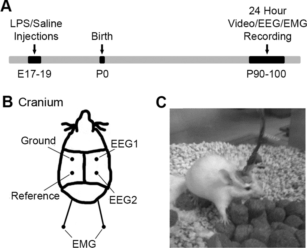
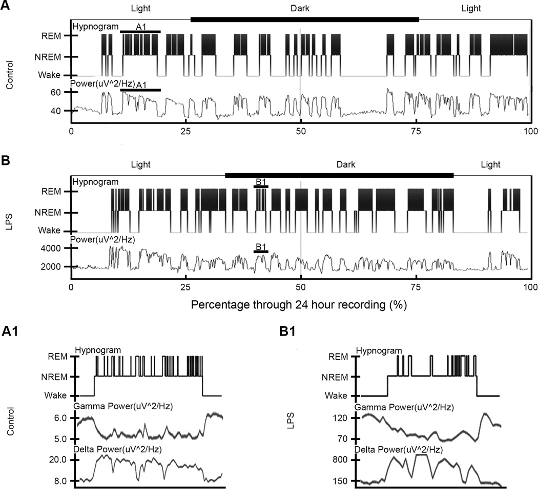
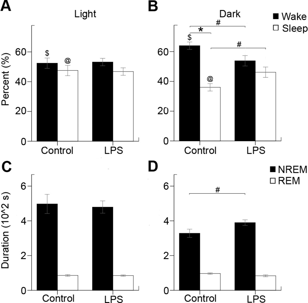
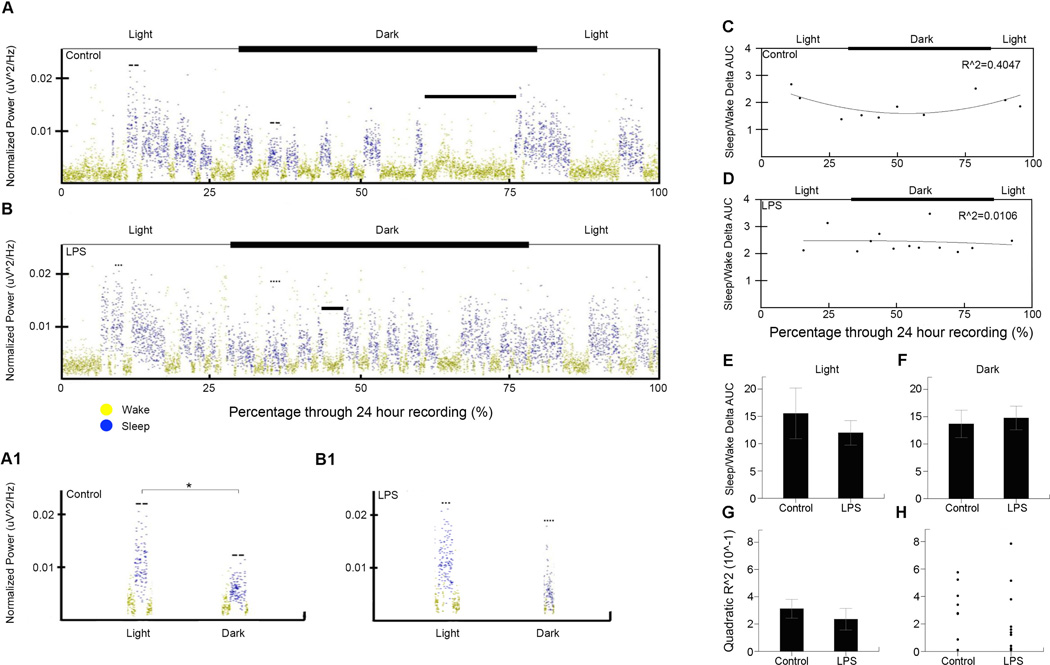
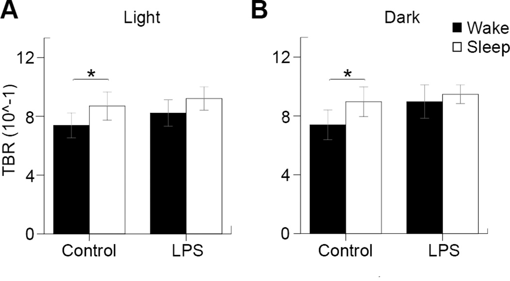
Intrauterine infection or inflammation in preterm neonates is a known risk for adverse neurological outcomes, including cognitive, motor and behavioral disabilities. Our previous data suggest that there is acute fetal brain inflammation in a mouse model of intrauterine exposure to lipopolysaccharides (LPS). We hypothesized that the in utero inflammation induced by LPS produces long-term electroencephalogram (EEG) biomarkers of neurodegeneration in the exposed mice that could be determined by using continuous quantitative video/EEG/electromyogram (EMG) analyses. A single LPS injection at E17 was performed in pregnant CD1 dams. Control dams were injected with same volumes of saline (LPS n=10, Control n=8). At postnatal age of P90-100, 24-h synchronous video/EEG/EMG recordings were done using a tethered recording system and implanted subdural electrodes. Behavioral state scoring was performed blind to treatment group, on each 10s EEG epoch using synchronous video, EMG and EEG trace signatures to generate individual hypnograms. Automated EEG power spectrums were analyzed for delta and theta-beta power ratios during wake vs. sleep cycles. Both control and LPS hypnograms showed an ultradian wake/sleep cycling. Since rodents are nocturnal animals, control mice showed the expected diurnal variation with significantly longer time spent in wake states during the dark cycle phase. In contrast, the LPS-treated mice lost this circadian rhythm. Sleep microstructure also showed significant alteration in the LPS mice specifically during the dark cycle, caused by significantly longer average non-rapid eye movement (NREM) cycle durations. No significance was found between treatment groups for the delta power data; however, significant activity-dependent changes in theta-beta power ratios seen in controls were absent in the LPS-exposed mice. In conclusion, exposure to in utero inflammation in CD1 mice resulted in significantly altered sleep architecture as adults that were circadian cycle and activity state dependent.
早产新生儿的宫内感染或炎症是导致不良神经学后果的已知风险因素,这些后果包括认知、运动和行为障碍。我们之前的数据表明,在宫内暴露于脂多糖(LPS)的小鼠模型中存在急性胎儿脑部炎症。我们假设,LPS诱导的宫内炎症会在暴露的小鼠中产生神经退行性变的长期脑电图(EEG)生物标志物,这可以通过连续定量视频/EEG/肌电图(EMG)分析来确定。在怀孕的CD1母鼠孕期第17天进行单次LPS注射。对照母鼠注射相同体积的生理盐水(LPS组n = 10,对照组n = 8)。在出生后第90 - 100天,使用系留记录系统和植入的硬膜下电极进行24小时同步视频/EEG/EMG记录。对治疗组进行盲法行为状态评分,在每个10秒的EEG时段使用同步视频、EMG和EEG波形特征生成个体睡眠图。在清醒与睡眠周期中分析自动EEG功率谱的δ和θ-β功率比。对照和LPS睡眠图均显示出超日节律性清醒/睡眠循环。由于啮齿动物是夜行性动物,对照小鼠表现出预期的昼夜变化,在黑暗周期阶段清醒状态下花费的时间明显更长。相比之下,LPS处理的小鼠失去了这种昼夜节律。睡眠微观结构在LPS小鼠中也显示出显著改变,特别是在黑暗周期,这是由平均非快速眼动(NREM)周期持续时间显著延长引起的。在治疗组之间,δ功率数据没有显著差异;然而,在LPS暴露的小鼠中没有观察到对照组中出现的与活动相关的θ-β功率比的显著变化。总之,CD1小鼠宫内炎症暴露导致成年后睡眠结构显著改变,这种改变依赖于昼夜节律周期和活动状态。