• 文献检索
  • 文档翻译
  • 深度研究
  • 学术资讯
  • Suppr Zotero 插件Zotero 插件
  • 邀请有礼
  • 套餐&价格
  • 历史记录
应用&插件
Suppr Zotero 插件Zotero 插件浏览器插件Mac 客户端Windows 客户端微信小程序
定价
高级版会员购买积分包购买API积分包
服务
文献检索文档翻译深度研究API 文档MCP 服务
关于我们
关于 Suppr公司介绍联系我们用户协议隐私条款
关注我们

Suppr 超能文献

核心技术专利:CN118964589B侵权必究
粤ICP备2023148730 号-1Suppr @ 2026

文献检索

告别复杂PubMed语法,用中文像聊天一样搜索,搜遍4000万医学文献。AI智能推荐,让科研检索更轻松。

立即免费搜索

文件翻译

保留排版,准确专业,支持PDF/Word/PPT等文件格式,支持 12+语言互译。

免费翻译文档

深度研究

AI帮你快速写综述,25分钟生成高质量综述,智能提取关键信息,辅助科研写作。

立即免费体验

计算机头影测量程序软组织治疗模拟模块的评估

Evaluation of the soft tissue treatment simulation module of a computerized cephalometric program.

作者信息

Oz Aslihan Zeynep, Akcan Cenk Ahmet, El Hakan, Ciger Semra

机构信息

Department of Orthodontics, Faculty of Dentistry, Ondokuz Mayis University, Samsun, Turkiye.

Department of Orthodontics, Faculty of Dentistry, Hacettepe University, Ankara, Turkiye.

出版信息

Eur J Dent. 2014 Apr;8(2):229-233. doi: 10.4103/1305-7456.130614.

DOI:10.4103/1305-7456.130614
PMID:24966775
原文链接:https://pmc.ncbi.nlm.nih.gov/articles/PMC4054055/
Abstract

OBJECTIVE

The purpose of this study is to compare the accuracy of the treatment simulation module of Quick Ceph Studio (QCS) program to the actual treatment results in Class II Division 1 patients.

DESIGN

Retrospective study.

MATERIALS AND METHODS

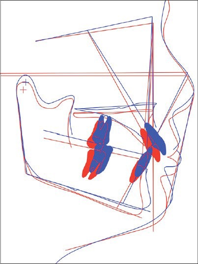
Twenty-six skeletal Class II patients treated with functional appliances were included. T0 and T1 lateral cephalograms were digitized using QCS. Before applying treatment simulation to the digitized cephalograms, the actual T0-T1 difference was calculated for the SNA, SNB, ANB angles, maxillary incisor inclination, and protrusion and mandibular incisor inclination and protrusion values. Next, using the treatment simulation module, the aforementioned values for the T0 cephalograms were manually entered to match the actual T1 values taking into account the T0-T1 differences. Paired sample t-test were applied to determine the difference between actual and treatment simulation measurements.

RESULTS

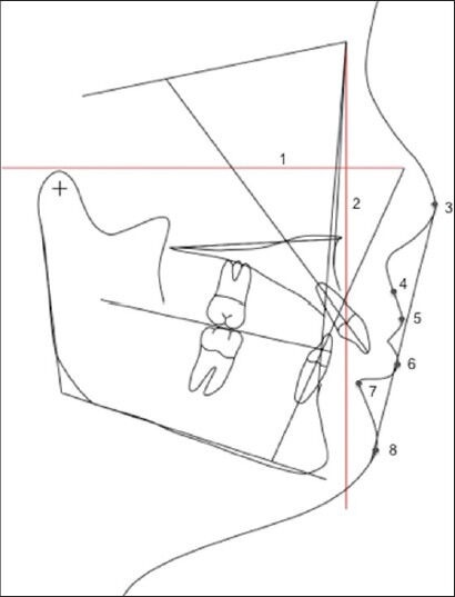
No significant differences were found for the anteroposterior location of the landmarks. Upper lip, soft tissue A point, soft tissue pogonion, and soft tissue B point measurements showed statistically significant difference between actual and treatment simulation in the vertical plane.

CONCLUSION

Quick Ceph program was reliable in terms of reflecting the sagittal changes that would probably occur with treatment and growth. However, vertical positions of the upper lip, soft tissue pogonion, soft tissue A point, and soft tissue B point were statistically different from actual results.

摘要

目的

本研究旨在比较Quick Ceph Studio(QCS)程序的治疗模拟模块与安氏II类1分类患者实际治疗结果的准确性。

设计

回顾性研究。

材料与方法

纳入26例使用功能矫治器治疗的骨骼型安氏II类患者。使用QCS对T0和T1侧位头影测量片进行数字化处理。在对数字化头影测量片应用治疗模拟之前,计算SNA、SNB、ANB角、上颌切牙倾斜度和突度以及下颌切牙倾斜度和突度值的实际T0 - T1差值。接下来,使用治疗模拟模块,考虑T0 - T1差值,手动输入T0头影测量片的上述值以匹配实际T1值。应用配对样本t检验来确定实际测量值与治疗模拟测量值之间的差异。

结果

在标志点的前后位置上未发现显著差异。上唇、软组织A点、软组织颏前点和软组织B点的测量在垂直平面上实际测量值与治疗模拟值之间存在统计学显著差异。

结论

Quick Ceph程序在反映治疗和生长可能发生的矢状面变化方面是可靠的。然而,上唇、软组织颏前点、软组织A点和软组织B点的垂直位置与实际结果在统计学上存在差异。

https://cdn.ncbi.nlm.nih.gov/pmc/blobs/4e3f/4054055/3b8eddbe5a75/EJD-8-229-g002.jpg
https://cdn.ncbi.nlm.nih.gov/pmc/blobs/4e3f/4054055/dc8bf61d139f/EJD-8-229-g001.jpg
https://cdn.ncbi.nlm.nih.gov/pmc/blobs/4e3f/4054055/3b8eddbe5a75/EJD-8-229-g002.jpg
https://cdn.ncbi.nlm.nih.gov/pmc/blobs/4e3f/4054055/dc8bf61d139f/EJD-8-229-g001.jpg
https://cdn.ncbi.nlm.nih.gov/pmc/blobs/4e3f/4054055/3b8eddbe5a75/EJD-8-229-g002.jpg

相似文献

1
Evaluation of the soft tissue treatment simulation module of a computerized cephalometric program.计算机头影测量程序软组织治疗模拟模块的评估
Eur J Dent. 2014 Apr;8(2):229-233. doi: 10.4103/1305-7456.130614.
2
Soft and hard tissue changes after bimaxillary surgery in Turkish female Class III patients.土耳其女性III类错颌患者双颌手术后的软硬组织变化
J Craniomaxillofac Surg. 2009 Jan;37(1):8-17. doi: 10.1016/j.jcms.2008.07.004. Epub 2008 Sep 10.
3
Evaluation of mandibular hard and soft tissues in cleft patients.腭裂患者下颌骨硬组织和软组织的评估。
J Istanb Univ Fac Dent. 2017 Apr 3;51(2):14-22. doi: 10.17096/jiufd.95754. eCollection 2017.
4
Customized maxillary incisor position relative to dentoskeletal and soft tissue patterns in Chinese women: A retrospective study.中国女性中定制的上颌切牙相对于牙颌骨及软组织形态的位置:一项回顾性研究。
Korean J Orthod. 2022 Mar 25;52(2):150-160. doi: 10.4041/kjod.2022.52.2.150.
5
Factors influencing soft tissue profile changes following orthodontic treatment in patients with Class II Division 1 malocclusion.安氏Ⅱ类1分类错牙合畸形患者正畸治疗后影响软组织侧貌变化的因素
Prog Orthod. 2016;17:13. doi: 10.1186/s40510-016-0125-1. Epub 2016 May 2.
6
Effects of activator and high-pull headgear combination therapy: skeletal, dentoalveolar, and soft tissue profile changes.激活器与高位牵引头帽联合治疗的效果:骨骼、牙牙槽及软组织侧貌变化
Eur J Orthod. 2007 Apr;29(2):140-8. doi: 10.1093/ejo/cjm003.
7
The effectiveness of pendulum, K-loop, and distal jet distalization techniques in growing children and its effects on anchor unit: A comparative study.钟摆式、K环和远中喷射远中移动技术在儿童生长发育中的有效性及其对支抗单位的影响:一项比较研究。
J Indian Soc Pedod Prev Dent. 2016 Oct-Dec;34(4):331-40. doi: 10.4103/0970-4388.191411.
8
Soft tissue and dentoskeletal profile changes associated with maxillary expansion and protraction headgear treatment.与上颌扩弓和口外牵引矫治器治疗相关的软组织和牙颌面形态变化。
Am J Orthod Dentofacial Orthop. 1996 Jan;109(1):38-49. doi: 10.1016/s0889-5406(96)70161-0.
9
Mandibular setback osteotomy: facial soft tissue behavior and possibility to improve the accuracy of the soft tissue profile prediction with the use of a computerized cephalometric program: Quick Ceph Image Pro: v. 2.5.下颌后缩截骨术:面部软组织行为以及使用计算机化头影测量程序(Quick Ceph Image Pro:v. 2.5)提高软组织侧貌预测准确性的可能性
Clin Orthod Res. 1999 May;2(2):85-98. doi: 10.1111/ocr.1999.2.2.85.
10
Evaluation of Soft Tissue Changes Around the Lips After Mandibular Setback Surgery With Minimal Orthodontics Using Three-Dimensional Stereophotogrammetry.使用三维立体摄影测量法评估下颌后缩手术联合微正畸治疗后唇部周围软组织的变化。
J Oral Maxillofac Surg. 2016 May;74(5):1044-54. doi: 10.1016/j.joms.2015.11.023. Epub 2015 Nov 24.

引用本文的文献

1
Which of the nine facial profiles according to AM Schwarz is the most (un)attractive?根据AM·施瓦茨的理论,九种面部轮廓中哪一种最具(不具)吸引力?
J Orofac Orthop. 2025 Apr 8. doi: 10.1007/s00056-025-00584-z.
2
Accuracy and reliability of Keynote for tracing and analyzing cephalometric radiographs.用于头影测量X线片追踪与分析的Keynote软件的准确性和可靠性
Angle Orthod. 2025 Mar 18;95(4):371-8. doi: 10.2319/101724-864.1.
3
Reliability of Photogrammetric Landmarks to the Conventional Cephalogram for Analyzing Soft-Tissue Landmarks in Orthodontics.

本文引用的文献

1
Prevalence of malocclusion and orthodontic treatment needs among 12-15 years old school children of Udaipur, India.印度乌代布尔12至15岁在校儿童错牙合畸形患病率及正畸治疗需求
Eur J Dent. 2013 Sep;7(Suppl 1):S045-S053. doi: 10.4103/1305-7456.119071.
2
Statistical power analyses using G*Power 3.1: tests for correlation and regression analyses.使用 G*Power 3.1 进行统计功效分析:相关和回归分析的检验。
Behav Res Methods. 2009 Nov;41(4):1149-60. doi: 10.3758/BRM.41.4.1149.
3
The accuracy of computer-assisted surgical planning in soft tissue prediction following orthognathic surgery.
用于正畸学中分析软组织标志点的摄影测量标志点相对于传统头影测量的可靠性
J Pharm Bioallied Sci. 2021 Jun;13(Suppl 1):S171-S175. doi: 10.4103/jpbs.JPBS_634_20. Epub 2021 Jun 5.
4
Full-mouth rehabilitation of Class II deep-bite patient: A 5-year clinical report.II类深覆合患者的全口修复:一份5年的临床报告。
Eur J Dent. 2016 Jul-Sep;10(3):426-431. doi: 10.4103/1305-7456.184160.
5
Determination of vertical characteristics with different cephalometric measurements.通过不同的头影测量法确定垂直特征。
Eur J Dent. 2016 Jan-Mar;10(1):116-120. doi: 10.4103/1305-7456.175694.
6
Pleasantness of facial profile and its correlation with soft tissue cephalometric parameters: Perception of orthodontists and lay people.面部侧貌的美观度及其与软组织头影测量参数的相关性:正畸医生和普通民众的认知
Eur J Dent. 2015 Jul-Sep;9(3):352-355. doi: 10.4103/1305-7456.163323.
正颌手术后软组织预测中计算机辅助手术规划的准确性。
Int J Med Robot. 2007 Mar;3:64-71. doi: 10.1002/rcs.127.
4
Accuracy of a LeFort I maxillary osteotomy.LeFort I型上颌骨截骨术的准确性。
Angle Orthod. 2005 Nov;75(6):964-73. doi: 10.1043/0003-3219(2005)75[964:AOALIM]2.0.CO;2.
5
Predictability of soft tissue profile changes following bimaxillary surgery in skeletal class III Chinese patients.骨性III类中国患者双颌手术后软组织侧貌变化的可预测性
J Oral Maxillofac Surg. 2004 Dec;62(12):1505-9. doi: 10.1016/j.joms.2004.04.022.
6
A comparison of current prediction imaging programs.当前预测成像程序的比较。
Am J Orthod Dentofacial Orthop. 2004 May;125(5):527-36. doi: 10.1016/S0889540604001210.
7
The accuracy of video imaging prediction in soft tissue outcome after bimaxillary orthognathic surgery.双颌正颌手术后软组织结果的视频成像预测准确性
J Oral Maxillofac Surg. 2003 Mar;61(3):333-42. doi: 10.1053/joms.2003.50058.
8
A radiographic analysis of computer prediction in conjunction with orthognathic surgery.计算机预测结合正颌外科手术的影像学分析
Int J Oral Maxillofac Surg. 2001 Aug;30(4):259-63. doi: 10.1054/ijom.2001.0089.
9
Accuracy of video imaging for predicting the soft tissue profile after mandibular set-back surgery.
Am J Orthod Dentofacial Orthop. 1999 Apr;115(4):382-9. doi: 10.1016/s0889-5406(99)70256-8.
10
Accuracy of cephalometric and video imaging program Dentofacial Planner Plus in orthognathic surgical planning.头影测量和视频成像程序Dentofacial Planner Plus在正颌外科手术规划中的准确性。
Comput Aided Surg. 1998;3(3):108-14. doi: 10.1002/(SICI)1097-0150(1998)3:3<108::AID-IGS2>3.0.CO;2-T.