Turunen Aaro, Hukkanen Veijo, Nygårdas Michaela, Kulmala Jarmo, Syrjänen Stina
Institute of Dentistry, Department of Oral Pathology, University of Turku, Lemminkäisenkatu 2, 20520 Turku, Finland.
Virol J. 2014 Jul 8;11:125. doi: 10.1186/1743-422X-11-125.
Oral mucosa is frequently exposed to Herpes simplex virus type 1 (HSV-1) infection and irradiation due to dental radiography. During radiotherapy for oral cancer, the surrounding clinically normal tissues are also irradiated. This prompted us to study the effects of HSV-1 infection and irradiation on viability and apoptosis of oral epithelial cells.
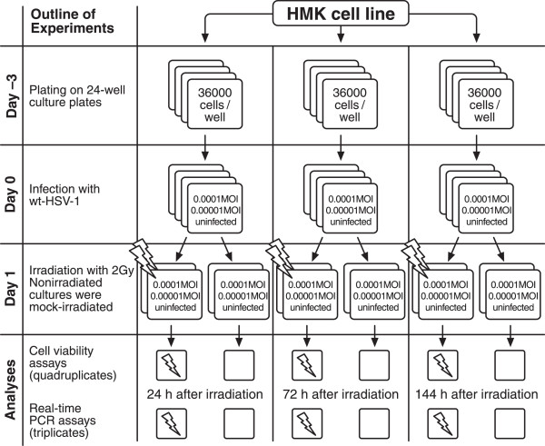
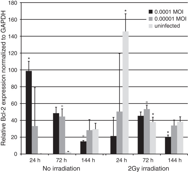
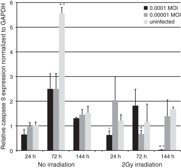
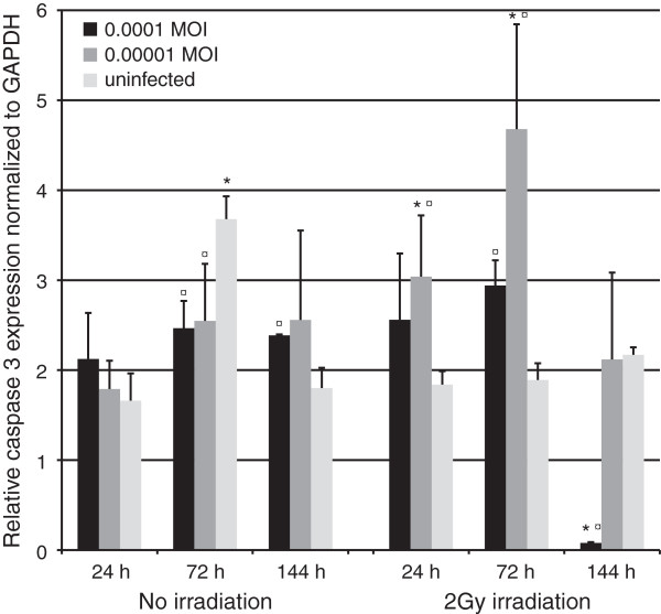
Immortal gingival keratinocyte (HMK) cells were infected with HSV-1 at a low multiplicity of infection (MOI) and irradiated with 2 Gy 24 hours post infection. The cells were then harvested at 24, 72 and 144 hours post irradiation for viability assays and qRT-PCR analyses for the apoptosis-related genes caspases 3, 8, and 9, bcl-2, NFκB1, and viral gene VP16. Mann-Whitney U-test was used for statistical calculations.
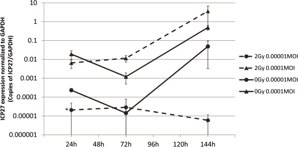
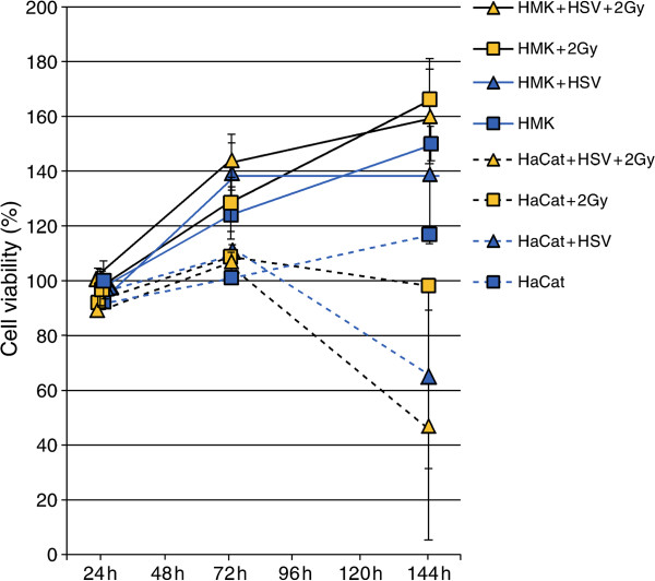
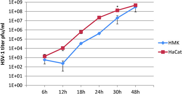
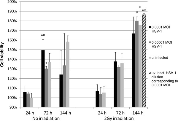
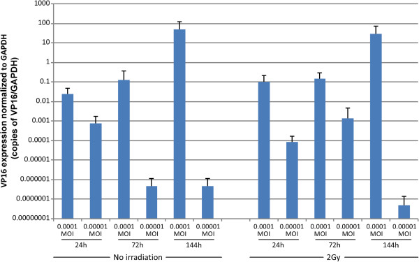
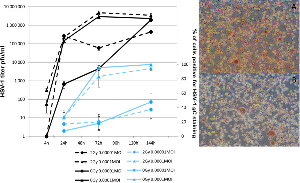
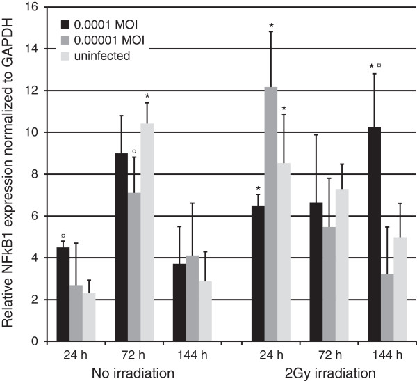
Irradiation improved the cell viability at 144 hours post irradiation (P = 0.05), which was further improved by HSV-1 infection at MOI of 0.00001 (P = 0.05). Simultaneously, the combined effects of infection at MOI of 0.0001 and irradiation resulted in upregulation in NFκB1 (P = 0.05). The combined effects of irradiation and HSV infection also significantly downregulated the expression of caspases 3, 8, and 9 at 144 hours (P = 0.05) whereas caspase 3 and 8 significantly upregulated in non-irradiated, HSV-infected cells as compared to uninfected controls (P = 0.05). Infection with 0.0001 MOI downregulated bcl-2 in non-irradiated cells but was upregulated by 27% after irradiation when compared to non-irradiated infected cells (P = 0.05). Irradiation had no effect on HSV-1 shedding or HSV gene expression at 144 hours.
HSV-1 infection may improve the viability of immortal cells after irradiation. The effect might be related to inhibition of apoptosis.
口腔黏膜经常因口腔X光检查而暴露于单纯疱疹病毒1型(HSV-1)感染和辐射中。在口腔癌放疗期间,周围临床正常组织也会受到辐射。这促使我们研究HSV-1感染和辐射对口腔上皮细胞活力和凋亡的影响。
永生化牙龈角质形成细胞(HMK)以低感染复数(MOI)感染HSV-1,并在感染后24小时接受2 Gy的辐射。然后在辐射后24、72和144小时收获细胞,进行活力测定以及对凋亡相关基因半胱天冬酶3、8和9、bcl-2、NFκB1和病毒基因VP16进行qRT-PCR分析。采用曼-惠特尼U检验进行统计计算。
辐射在辐射后144小时提高了细胞活力(P = 0.05),以0.00001的MOI进行HSV-1感染可进一步提高细胞活力(P = 0.05)。同时,以0.0001的MOI进行感染和辐射的联合作用导致NFκB1上调(P = 0.05)。辐射和HSV感染的联合作用在144小时时也显著下调了半胱天冬酶3、8和9的表达(P = 0.05),而与未感染对照相比,在未辐射、HSV感染的细胞中半胱天冬酶3和8显著上调(P = 0.05)。以0.0001的MOI进行感染在未辐射细胞中下调了bcl-2,但与未辐射感染细胞相比,辐射后上调了27%(P = 0.05)。辐射在144小时时对HSV-1脱落或HSV基因表达没有影响。
HSV-1感染可能提高辐射后永生化细胞的活力。这种作用可能与凋亡抑制有关。