Kalderstam Jonas, Sadik May, Edenbrandt Lars, Ohlsson Mattias
Department of Astronomy and Theoretical Physics, Lund University, Lund, Sweden.
BMC Med Imaging. 2014 Jul 10;14:24. doi: 10.1186/1471-2342-14-24.
A bone scan is a common method for monitoring bone metastases in patients with advanced prostate cancer. The Bone Scan Index (BSI) measures the tumor burden on the skeleton, expressed as a percentage of the total skeletal mass. Previous studies have shown that BSI is associated with survival of prostate cancer patients. The objective in this study was to investigate to what extent regional BSI measurements, as obtained by an automated method, can improve the survival analysis for advanced prostate cancer.
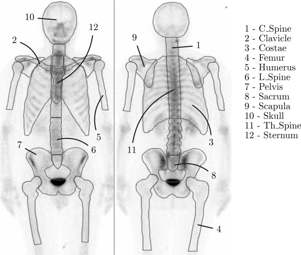
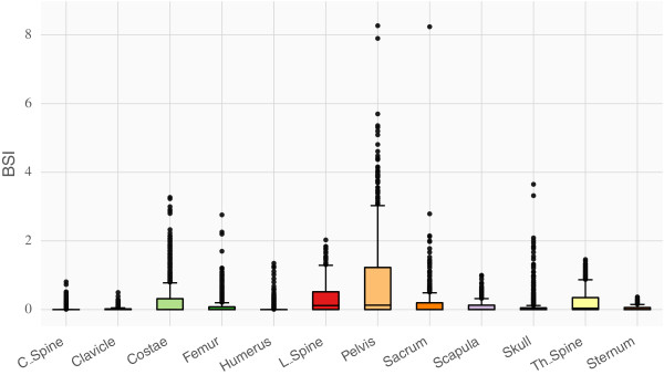
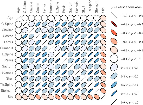
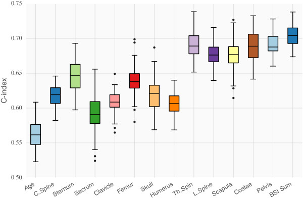
The automated method for analyzing bone scan images computed BSI values for twelve skeletal regions, in a study population consisting of 1013 patients diagnosed with prostate cancer. In the survival analysis we used the standard Cox proportional hazards model and a more advanced non-linear method based on artificial neural networks. The concordance index (C-index) was used to measure the performance of the models.
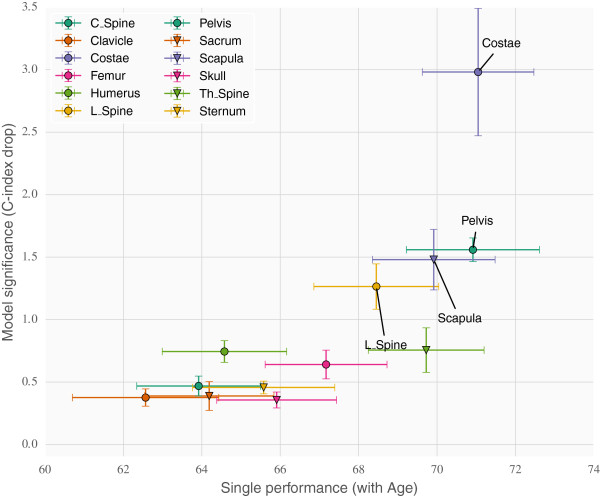
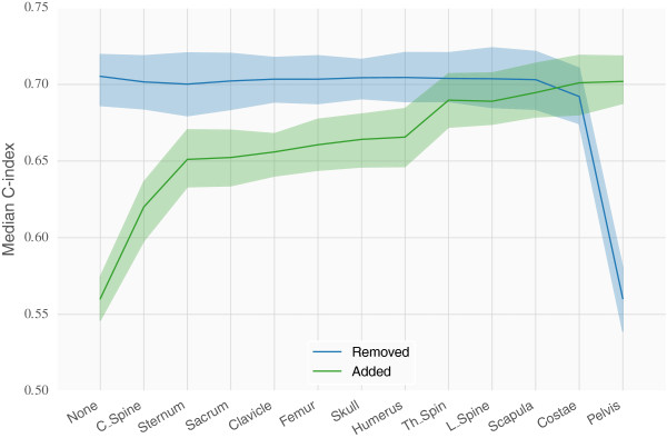
A Cox model with age and total BSI obtained a C-index of 70.4%. The best Cox model with regional measurements from Costae, Pelvis, Scapula and the Spine, together with age, got a similar C-index (70.5%). The overall best single skeletal localisation, as measured by the C-index, was Costae. The non-linear model performed equally well as the Cox model, ruling out any significant non-linear interactions among the regional BSI measurements.
The present study showed that the localisation of bone metastases obtained from the bone scans in prostate cancer patients does not improve the performance of the survival models compared to models using the total BSI. However a ranking procedure indicated that some regions are more important than others.
骨扫描是监测晚期前列腺癌患者骨转移的常用方法。骨扫描指数(BSI)衡量骨骼上的肿瘤负荷,以占骨骼总质量的百分比表示。先前的研究表明,BSI与前列腺癌患者的生存率相关。本研究的目的是调查通过自动方法获得的区域BSI测量在多大程度上可以改善晚期前列腺癌的生存分析。
在一个由1013名被诊断为前列腺癌的患者组成的研究群体中,用于分析骨扫描图像的自动方法计算了12个骨骼区域的BSI值。在生存分析中,我们使用了标准的Cox比例风险模型和基于人工神经网络的更先进的非线性方法。一致性指数(C指数)用于衡量模型的性能。
一个包含年龄和总BSI的Cox模型的C指数为70.4%。包含肋骨、骨盆、肩胛骨和脊柱的区域测量值以及年龄的最佳Cox模型得到了相似的C指数(70.5%)。以C指数衡量,总体最佳的单个骨骼定位是肋骨。非线性模型的表现与Cox模型相当,排除了区域BSI测量值之间任何显著的非线性相互作用。
本研究表明,与使用总BSI的模型相比,前列腺癌患者骨扫描中获得的骨转移定位并不能提高生存模型的性能。然而,一个排序程序表明某些区域比其他区域更重要。