• 文献检索
  • 文档翻译
  • 深度研究
  • 学术资讯
  • Suppr Zotero 插件Zotero 插件
  • 邀请有礼
  • 套餐&价格
  • 历史记录
应用&插件
Suppr Zotero 插件Zotero 插件浏览器插件Mac 客户端Windows 客户端微信小程序
定价
高级版会员购买积分包购买API积分包
服务
文献检索文档翻译深度研究API 文档MCP 服务
关于我们
关于 Suppr公司介绍联系我们用户协议隐私条款
关注我们

Suppr 超能文献

核心技术专利:CN118964589B侵权必究
粤ICP备2023148730 号-1Suppr @ 2026

文献检索

告别复杂PubMed语法,用中文像聊天一样搜索,搜遍4000万医学文献。AI智能推荐,让科研检索更轻松。

立即免费搜索

文件翻译

保留排版,准确专业,支持PDF/Word/PPT等文件格式,支持 12+语言互译。

免费翻译文档

深度研究

AI帮你快速写综述,25分钟生成高质量综述,智能提取关键信息,辅助科研写作。

立即免费体验

实验性青光眼对侧的小鼠视网膜中的小胶质细胞在所有视网膜层均表现出多种激活迹象。

Microglia in mouse retina contralateral to experimental glaucoma exhibit multiple signs of activation in all retinal layers.

作者信息

Rojas Blanca, Gallego Beatriz I, Ramírez Ana I, Salazar Juan J, de Hoz Rosa, Valiente-Soriano Francisco J, Avilés-Trigueros Marcelino, Villegas-Perez Maria P, Vidal-Sanz Manuel, Triviño Alberto, Ramírez José M

机构信息

Instituto de Investigaciones Oftalmológicas Ramón Castroviejo, Facultad de Medicina, Pab VI, 4a, Avenida Complutense s/n, Universidad Complutense de Madrid, 28040 Madrid, Spain.

出版信息

J Neuroinflammation. 2014 Jul 26;11:133. doi: 10.1186/1742-2094-11-133.

DOI:10.1186/1742-2094-11-133
PMID:25064005
原文链接:https://pmc.ncbi.nlm.nih.gov/articles/PMC4128533/
Abstract

BACKGROUND

Glaucomatous optic neuropathy, a leading cause of blindness, can progress despite control of intraocular pressure - currently the main risk factor and target for treatment. Glaucoma progression shares mechanisms with neurodegenerative disease, including microglia activation. In the present model of ocular hypertension (OHT), we have recently described morphological signs of retinal microglia activation and MHC-II upregulation in both the untreated contralateral eyes and OHT eyes. By using immunostaining, we sought to analyze and quantify additional signs of microglia activation and differences depending on the retinal layer.

METHODS

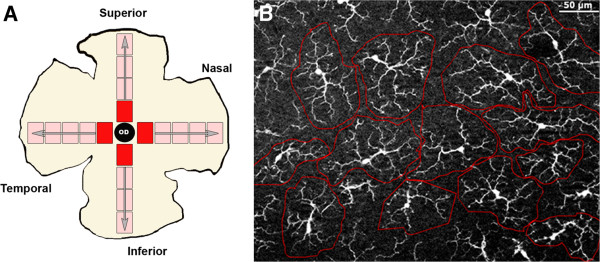
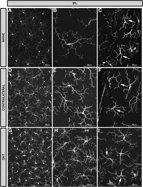
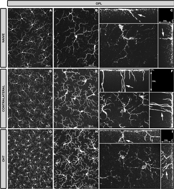
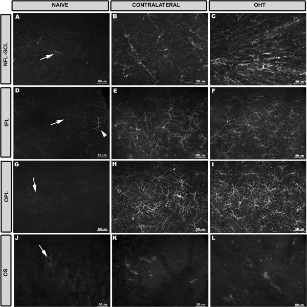
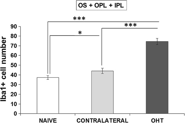
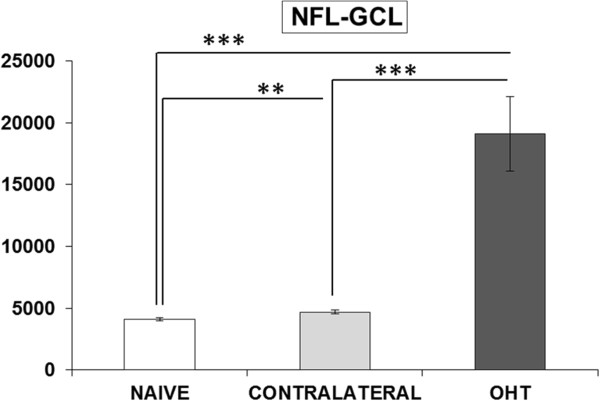
Two groups of adult Swiss mice were used: age-matched control (naïve, n = 12), and lasered (n = 12). In the lasered animals, both OHT eyes and contralateral eyes were analyzed. Retinal whole-mounts were immunostained with antibodies against Iba-1, MHC-II, CD68, CD86, and Ym1. The Iba-1+ cell number in the plexiform layers (PL) and the photoreceptor outer segment (OS), Iba-1+ arbor area in the PL, and area of the retina occupied by Iba-1+ cells in the nerve fiber layer-ganglion cell layer (NFL-GCL) were quantified.

RESULTS

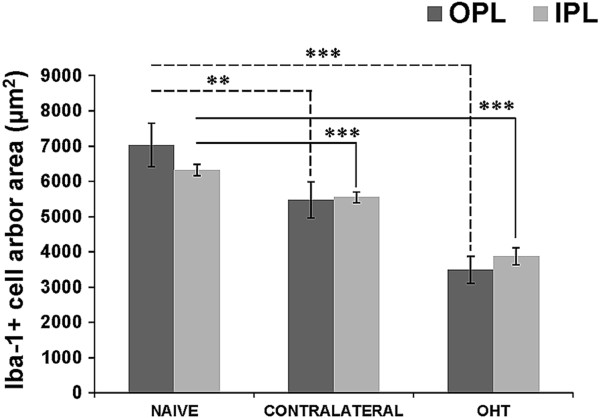
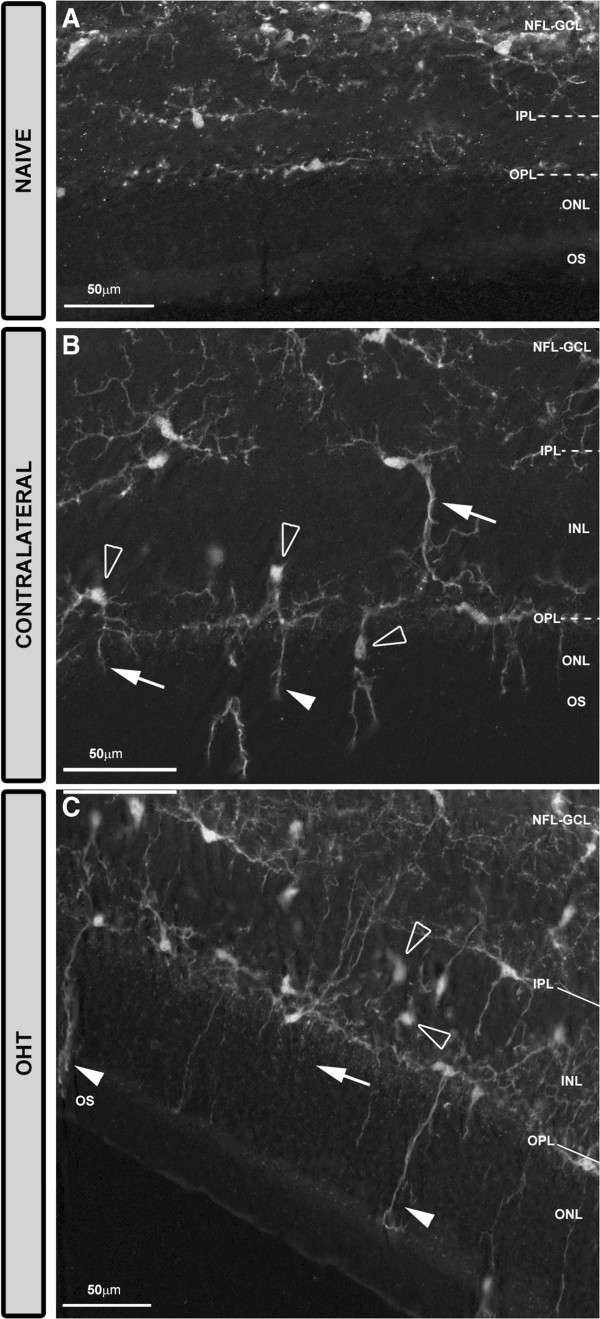
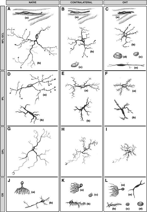
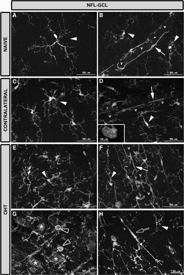
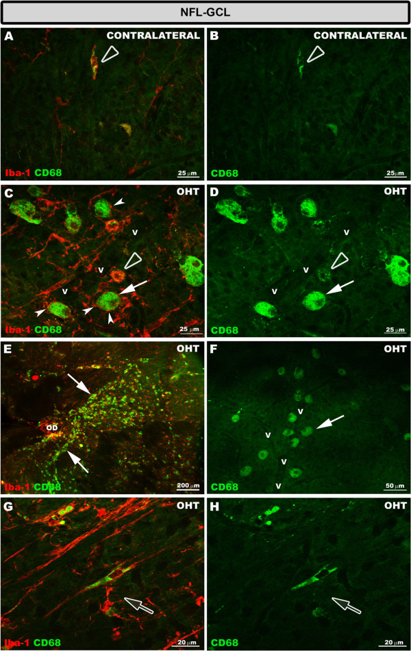
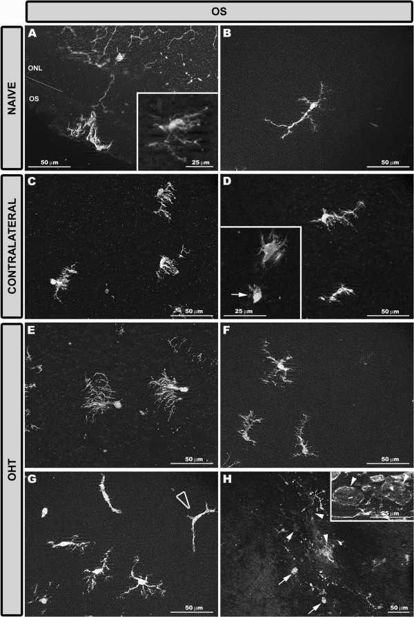
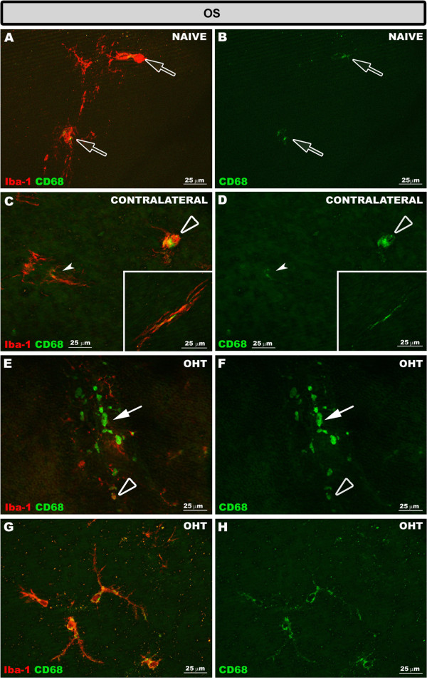
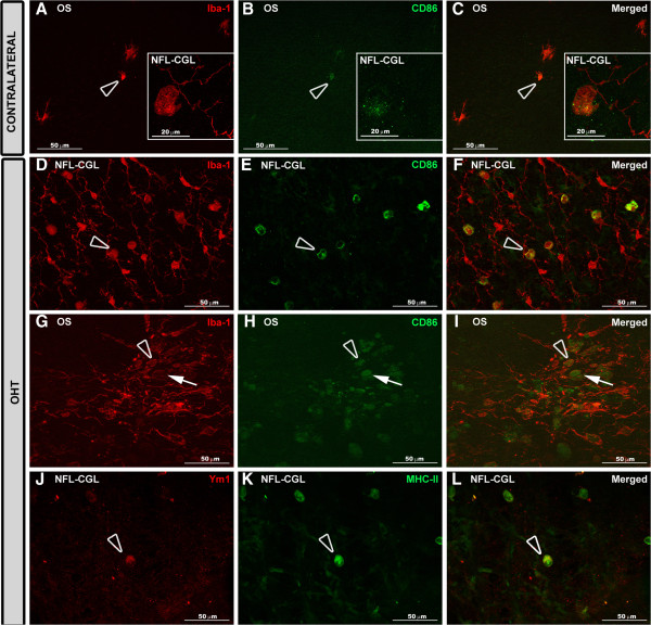
The main findings in contralateral eyes and OHT eyes were: i) ameboid microglia in the NFL-GCL and OS; ii) the retraction of processes in all retinal layers; iii) a higher level of branching in PL and in the OS; iv) soma displacement to the nearest cell layers in the PL and OS; v) the reorientation of processes in the OS; vi) MHC-II upregulation in all retinal layers; vii) increased CD68 immunostaining; and viii) CD86 immunolabeling in ameboid cells. In comparison with the control group, a significant increase in the microglial number in the PL, OS, and in the area occupied by Iba-1+ cells in the NFL-GCL, and significant reduction of the arbor area in the PL. In addition, rounded Iba-1+ CD86+ cells in the NFL-GCL, OS and Ym1+ cells, and rod-like microglia in the NFL-GCL were restricted to OHT eyes.

CONCLUSIONS

Several quantitative and qualitative signs of microglia activation are detected both in the contralateral and OHT eyes. Such activation extended beyond the GCL, involving all retinal layers. Differences between the two eyes could help to elucidate glaucoma pathophysiology.

摘要

背景

青光眼性视神经病变是失明的主要原因之一,尽管眼压得到控制(目前眼压是主要危险因素和治疗靶点),病情仍可能进展。青光眼进展与神经退行性疾病有共同机制,包括小胶质细胞激活。在当前的高眼压(OHT)模型中,我们最近描述了未治疗的对侧眼和高眼压眼中视网膜小胶质细胞激活的形态学迹象以及MHC-II上调。通过免疫染色,我们试图分析和量化小胶质细胞激活的其他迹象以及视网膜各层之间的差异。

方法

使用两组成年瑞士小鼠:年龄匹配的对照组(未处理,n = 12)和激光照射组(n = 12)。在激光照射的动物中,对高眼压眼和对侧眼均进行分析。视网膜全层标本用抗Iba-1、MHC-II、CD68、CD86和Ym1的抗体进行免疫染色。对视神经纤维层-神经节细胞层(NFL-GCL)中丛状层(PL)和光感受器外段(OS)的Iba-1+细胞数量、PL中Iba-1+树突面积以及Iba-1+细胞占据的视网膜面积进行量化。

结果

对侧眼和高眼压眼的主要发现如下:i)NFL-GCL和OS中有阿米巴样小胶质细胞;ii)所有视网膜层的突起回缩;iii)PL和OS中分支水平更高;iv)PL和OS中细胞体向最近的细胞层移位;v)OS中突起重新定向;vi)所有视网膜层中MHC-II上调;vii)CD68免疫染色增加;viii)阿米巴样细胞中有CD86免疫标记。与对照组相比,PL、OS以及NFL-GCL中Iba-1+细胞占据区域的小胶质细胞数量显著增加,PL中树突面积显著减少。此外,NFL-GCL、OS中的圆形Iba-1+ CD86+细胞和Ym1+细胞以及NFL-GCL中的杆状小胶质细胞仅限于高眼压眼。

结论

在对侧眼和高眼压眼中均检测到小胶质细胞激活的若干定量和定性迹象。这种激活超出了神经节细胞层,涉及所有视网膜层。两只眼睛之间的差异可能有助于阐明青光眼的病理生理学。

https://cdn.ncbi.nlm.nih.gov/pmc/blobs/5db9/4128533/75b8ff826237/1742-2094-11-133-14.jpg
https://cdn.ncbi.nlm.nih.gov/pmc/blobs/5db9/4128533/41e6d0686a1c/1742-2094-11-133-1.jpg
https://cdn.ncbi.nlm.nih.gov/pmc/blobs/5db9/4128533/f12cbc31ae21/1742-2094-11-133-2.jpg
https://cdn.ncbi.nlm.nih.gov/pmc/blobs/5db9/4128533/07db75080fa0/1742-2094-11-133-3.jpg
https://cdn.ncbi.nlm.nih.gov/pmc/blobs/5db9/4128533/85a85fcdf857/1742-2094-11-133-4.jpg
https://cdn.ncbi.nlm.nih.gov/pmc/blobs/5db9/4128533/6f397e843193/1742-2094-11-133-5.jpg
https://cdn.ncbi.nlm.nih.gov/pmc/blobs/5db9/4128533/abe5a472cd95/1742-2094-11-133-6.jpg
https://cdn.ncbi.nlm.nih.gov/pmc/blobs/5db9/4128533/fd7daf9a1c58/1742-2094-11-133-7.jpg
https://cdn.ncbi.nlm.nih.gov/pmc/blobs/5db9/4128533/eeb26f75f4fa/1742-2094-11-133-8.jpg
https://cdn.ncbi.nlm.nih.gov/pmc/blobs/5db9/4128533/021ef6c78f84/1742-2094-11-133-9.jpg
https://cdn.ncbi.nlm.nih.gov/pmc/blobs/5db9/4128533/6e0fb77bf75b/1742-2094-11-133-10.jpg
https://cdn.ncbi.nlm.nih.gov/pmc/blobs/5db9/4128533/7ad3456730eb/1742-2094-11-133-11.jpg
https://cdn.ncbi.nlm.nih.gov/pmc/blobs/5db9/4128533/7f3d568bf1e8/1742-2094-11-133-12.jpg
https://cdn.ncbi.nlm.nih.gov/pmc/blobs/5db9/4128533/eb311bca11a8/1742-2094-11-133-13.jpg
https://cdn.ncbi.nlm.nih.gov/pmc/blobs/5db9/4128533/75b8ff826237/1742-2094-11-133-14.jpg

相似文献

1
Microglia in mouse retina contralateral to experimental glaucoma exhibit multiple signs of activation in all retinal layers.实验性青光眼对侧的小鼠视网膜中的小胶质细胞在所有视网膜层均表现出多种激活迹象。
J Neuroinflammation. 2014 Jul 26;11:133. doi: 10.1186/1742-2094-11-133.
2
Bilateral early activation of retinal microglial cells in a mouse model of unilateral laser-induced experimental ocular hypertension.在单侧激光诱导实验性眼高压小鼠模型中观察到视网膜小胶质细胞的双侧早期激活。
Exp Eye Res. 2018 Jun;171:12-29. doi: 10.1016/j.exer.2018.03.006. Epub 2018 Mar 9.
3
Rod-like microglia are restricted to eyes with laser-induced ocular hypertension but absent from the microglial changes in the contralateral untreated eye.杆状小胶质细胞仅限于激光诱导性高眼压的眼睛,而对侧未治疗眼睛的小胶质细胞变化中则不存在。
PLoS One. 2013 Dec 18;8(12):e83733. doi: 10.1371/journal.pone.0083733. eCollection 2013.
4
IOP induces upregulation of GFAP and MHC-II and microglia reactivity in mice retina contralateral to experimental glaucoma.IOP 诱导实验性青光眼对侧小鼠视网膜 GFAP 和 MHC-II 的上调和小胶质细胞反应性。
J Neuroinflammation. 2012 May 14;9:92. doi: 10.1186/1742-2094-9-92.
5
Automatic Counting of Microglial Cells in Healthy and Glaucomatous Mouse Retinas.健康和青光眼小鼠视网膜中小胶质细胞的自动计数
PLoS One. 2015 Nov 18;10(11):e0143278. doi: 10.1371/journal.pone.0143278. eCollection 2015.
6
Time course of bilateral microglial activation in a mouse model of laser-induced glaucoma.激光诱导青光眼小鼠模型中双侧小胶质细胞激活的时程。
Sci Rep. 2020 Mar 17;10(1):4890. doi: 10.1038/s41598-020-61848-9.
7
Microglial changes in the early aging stage in a healthy retina and an experimental glaucoma model.健康视网膜和实验性青光眼模型中早期衰老阶段的小胶质细胞变化。
Prog Brain Res. 2020;256(1):125-149. doi: 10.1016/bs.pbr.2020.05.024. Epub 2020 Jun 16.
8
Inhibition of the cysteinyl leukotriene pathways increases survival of RGCs and reduces microglial activation in ocular hypertension.抑制半胱氨酰白三烯途径可增加 RGCs 的存活并减少眼高压中的小胶质细胞激活。
Exp Eye Res. 2021 Dec;213:108806. doi: 10.1016/j.exer.2021.108806. Epub 2021 Oct 27.
9
Time-dependent retinal ganglion cell loss, microglial activation and blood-retina-barrier tightness in an acute model of ocular hypertension.高眼压急性模型中视网膜神经节细胞的时间依赖性丢失、小胶质细胞激活及血视网膜屏障紧密性
Exp Eye Res. 2015 Jul;136:59-71. doi: 10.1016/j.exer.2015.05.010. Epub 2015 May 20.
10
Bilateral neuroinflammatory processes in visual pathways induced by unilateral ocular hypertension in the rat.大鼠单侧高眼压诱导的视觉通路双侧神经炎症过程。
J Neuroinflammation. 2016 Feb 20;13:44. doi: 10.1186/s12974-016-0509-7.

引用本文的文献

1
Tumor Necrosis Factor Alpha-Mediated Interaction Between Microglia and Müller Cells Exacerbates Retinal Ganglion Cell Damage in Experimental Glaucoma.肿瘤坏死因子α介导的小胶质细胞与米勒细胞之间的相互作用加剧实验性青光眼中的视网膜神经节细胞损伤
Neurosci Bull. 2025 Aug 30. doi: 10.1007/s12264-025-01478-1.
2
Ocular changes as potential biomarkers for early diagnosis of Alzheimer's disease.眼部变化作为阿尔茨海默病早期诊断的潜在生物标志物。
Alzheimers Dement. 2025 Aug;21(8):e70476. doi: 10.1002/alz.70476.
3
Effects of a ROCK Inhibitor on Retinal Ganglion Cells In Vivo and In Vitro.

本文引用的文献

1
Dynamic structural remodelling of microglia in health and disease: a review of the models, the signals and the mechanisms.健康和疾病状态下小胶质细胞的动态结构重塑:对模型、信号和机制的综述。
Brain Behav Immun. 2014 Mar;37:1-14. doi: 10.1016/j.bbi.2013.12.010. Epub 2014 Jan 8.
2
Rod-like microglia are restricted to eyes with laser-induced ocular hypertension but absent from the microglial changes in the contralateral untreated eye.杆状小胶质细胞仅限于激光诱导性高眼压的眼睛,而对侧未治疗眼睛的小胶质细胞变化中则不存在。
PLoS One. 2013 Dec 18;8(12):e83733. doi: 10.1371/journal.pone.0083733. eCollection 2013.
3
Acute and chronic stress-induced disturbances of microglial plasticity, phenotype and function.
ROCK抑制剂对视网膜神经节细胞的体内和体外作用
J Clin Med. 2025 Jul 29;14(15):5344. doi: 10.3390/jcm14155344.
4
SUMOylation is a Translatable Target in Hypoxic MNPs Regulating Retinal Vasculopathy.SUMO化修饰是调节视网膜血管病变的缺氧微小神经胶质细胞中的一个可翻译靶点。
Adv Sci (Weinh). 2025 Aug;12(31):e03505. doi: 10.1002/advs.202503505. Epub 2025 May 31.
5
Microglia remodeling in the visual thalamus of the DBA/2J mouse model of glaucoma.青光眼DBA/2J小鼠模型视丘中小胶质细胞的重塑
PLoS One. 2025 May 15;20(5):e0323513. doi: 10.1371/journal.pone.0323513. eCollection 2025.
6
Cannabinoids as Multitarget Drugs for the Treatment of Autoimmunity in Glaucoma.大麻素作为治疗青光眼自身免疫性疾病的多靶点药物。
ACS Pharmacol Transl Sci. 2025 Mar 26;8(4):932-950. doi: 10.1021/acsptsci.4c00583. eCollection 2025 Apr 11.
7
Targeting Connexin 43 in Retinal Astrocytes Promotes Neuronal Survival in Glaucomatous Injury.靶向视网膜星形胶质细胞中的连接蛋白43可促进青光眼性损伤中的神经元存活。
Glia. 2025 Jul;73(7):1398-1419. doi: 10.1002/glia.70013. Epub 2025 Mar 28.
8
Optic Nerve Crush Does Not Induce Retinal Ganglion Cell Loss in the Contralateral Eye.视神经挤压不会导致对侧眼视网膜神经节细胞丢失。
Invest Ophthalmol Vis Sci. 2025 Mar 3;66(3):49. doi: 10.1167/iovs.66.3.49.
9
Loss of monocyte chemoattractant protein-1 reduced monocyte recruitment and preserved retinal ganglion cells in a mouse model of hypertensive glaucoma.在高血压性青光眼小鼠模型中,单核细胞趋化蛋白-1的缺失减少了单核细胞募集并保护了视网膜神经节细胞。
Exp Eye Res. 2025 May;254:110325. doi: 10.1016/j.exer.2025.110325. Epub 2025 Mar 7.
10
Damage and repair in retinal degenerative diseases: Molecular basis through clinical translation.视网膜退行性疾病中的损伤与修复:从分子基础到临床转化
Neural Regen Res. 2026 Apr 1;21(4):1383-1395. doi: 10.4103/NRR.NRR-D-24-01016. Epub 2025 Feb 24.
急性和慢性应激引起的小胶质细胞可塑性、表型和功能障碍。
Curr Drug Targets. 2013 Oct;14(11):1262-76. doi: 10.2174/13894501113149990208.
4
Enhanced insight into the autoimmune component of glaucoma: IgG autoantibody accumulation and pro-inflammatory conditions in human glaucomatous retina.增强对青光眼自身免疫成分的认识:人青光眼视网膜中 IgG 自身抗体的积累和促炎状态。
PLoS One. 2013;8(2):e57557. doi: 10.1371/journal.pone.0057557. Epub 2013 Feb 25.
5
A quantitative spatiotemporal analysis of microglia morphology during ischemic stroke and reperfusion.定量时空分析缺血性脑卒中及再灌注后小胶质细胞形态变化。
J Neuroinflammation. 2013 Jan 11;10:4. doi: 10.1186/1742-2094-10-4.
6
Effect of brain-derived neurotrophic factor on mouse axotomized retinal ganglion cells and phagocytic microglia.脑源性神经营养因子对小鼠视神经切断后视网膜神经节细胞和吞噬性小胶质细胞的影响。
Invest Ophthalmol Vis Sci. 2013 Feb 1;54(2):974-85. doi: 10.1167/iovs.12-11207.
7
Morphological and genetic activation of microglia after diffuse traumatic brain injury in the rat.大鼠弥漫性创伤性脑损伤后小胶质细胞的形态和遗传激活。
Neuroscience. 2012 Dec 6;225:65-75. doi: 10.1016/j.neuroscience.2012.08.058. Epub 2012 Sep 6.
8
Early reduction of microglia activation by irradiation in a model of chronic glaucoma.照射对慢性青光眼模型中小胶质细胞激活的早期抑制作用。
PLoS One. 2012;7(8):e43602. doi: 10.1371/journal.pone.0043602. Epub 2012 Aug 30.
9
Microglial activation in the injured and healthy brain: what are we really talking about? Practical and theoretical issues associated with the measurement of changes in microglial morphology.损伤和健康大脑中的小胶质细胞激活:我们到底在谈论什么?与测量小胶质细胞形态变化相关的实际和理论问题。
Neuroscience. 2012 Dec 6;225:162-71. doi: 10.1016/j.neuroscience.2012.07.029. Epub 2012 Jul 20.
10
IOP induces upregulation of GFAP and MHC-II and microglia reactivity in mice retina contralateral to experimental glaucoma.IOP 诱导实验性青光眼对侧小鼠视网膜 GFAP 和 MHC-II 的上调和小胶质细胞反应性。
J Neuroinflammation. 2012 May 14;9:92. doi: 10.1186/1742-2094-9-92.