Instituto de Investigaciones Oftalmológicas Ramón Castroviejo, Universidad Complutense de Madrid, Madrid, Spain.
Facultad de Óptica y Optometría, Departamento de Inmunología, Oftalmología y ORL, Universidad Complutense de Madrid, Madrid, Spain.
Sci Rep. 2020 Mar 17;10(1):4890. doi: 10.1038/s41598-020-61848-9.
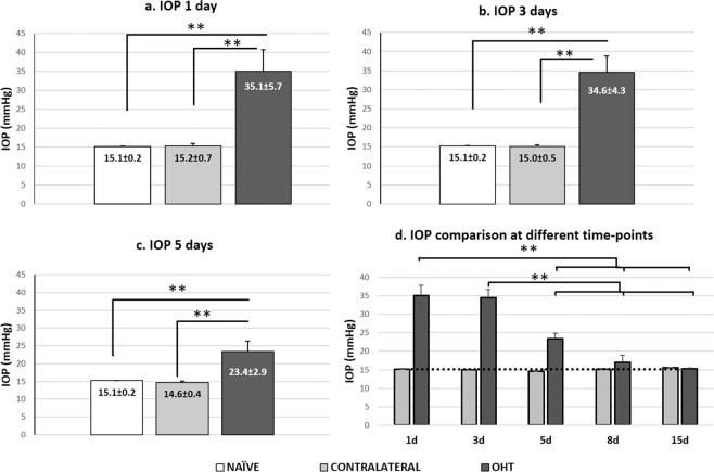
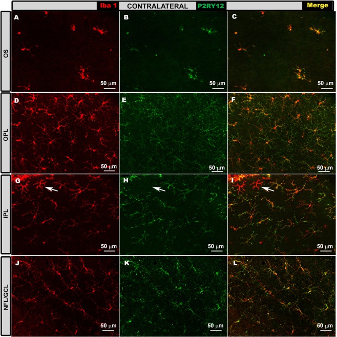
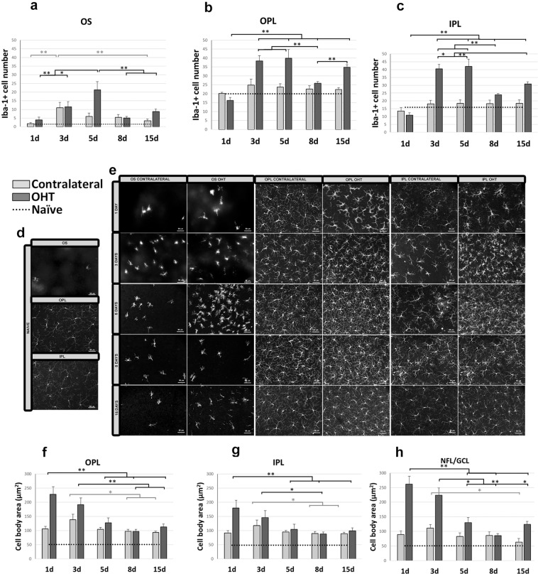
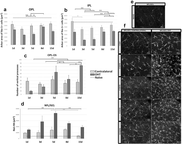
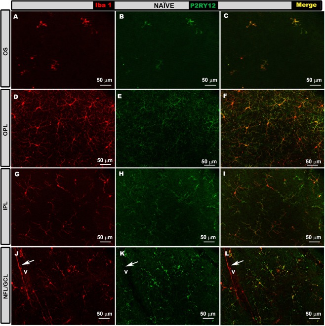
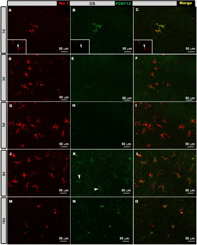
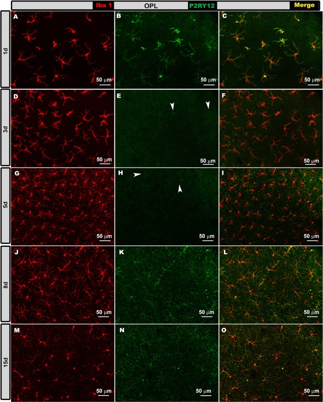
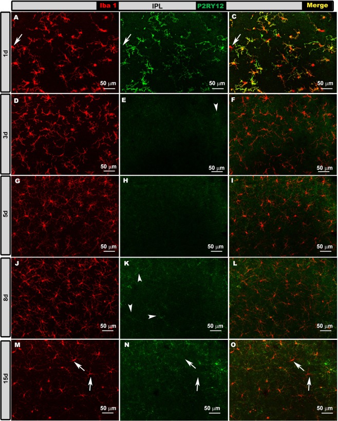
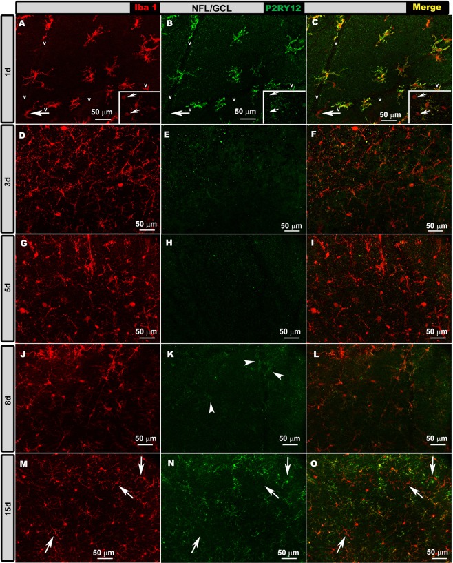
Microglial activation is associated with glaucoma. In the model of unilateral laser-induced ocular hypertension (OHT), the time point at which the inflammatory process peaks remains unknown. Different time points (1, 3, 5, 8, and 15 d) were compared to analyze signs of microglial activation both in OHT and contralateral eyes. In both eyes, microglial activation was detected in all retinal layers at all time points analyzed, including: i) increase in the cell number in the outer segment photoreceptor layer and plexiform layers (only in OHT eyes) from 3 d onward; ii) increase in soma size from 1 d onward; iii) retraction of the processes from 1 d in OHT eyes and 3 d in contralateral eyes; iv) increase in the area of the retina occupied by Iba-1+ cells in the nerve fiber layer/ganglion cell layer from 1 d onward; v) increase in the number of vertical processes from 1 d in contralateral eyes and 3 d in OHT eyes. In OHT eyes at 24 h and 15 d, most Iba-1+ cells were P2RY12+ and were down-regulated at 3 and 5 d. In both eyes, microglial activation was stronger at 3 and 5 d (inflammation peaked in this model). These time points could be useful to identify factors implicated in the inflammatory process.
小胶质细胞激活与青光眼有关。在单侧激光诱导的眼高压(OHT)模型中,炎症过程的峰值时间点尚不清楚。比较了不同时间点(1、3、5、8 和 15 天),以分析 OHT 和对侧眼中小胶质细胞激活的迹象。在所有分析的时间点,包括:i)在 3 天后,外节光感受器层和丛状层中的细胞数量增加(仅在 OHT 眼中);ii)从 1 天开始,体大小增加;iii)从 1 天开始,OHT 眼中的过程回缩,对侧眼中的过程从 3 天开始回缩;iv)从 1 天开始,神经纤维层/节细胞层中 Iba-1+细胞占据的视网膜面积增加;v)从 1 天开始,对侧眼中的垂直过程数量增加,OHT 眼中的垂直过程数量从 3 天开始增加。在 OHT 眼中,24 小时和 15 天,大多数 Iba-1+细胞是 P2RY12+,在 3 天和 5 天被下调。在两只眼中,3 天和 5 天的小胶质细胞激活更强(该模型中的炎症达到峰值)。这些时间点可能有助于识别炎症过程中涉及的因素。