Buchmann Andreas, Dentico Daniela, Peterson Michael J, Riedner Brady A, Sarasso Simone, Massimini Marcello, Tononi Giulio, Ferrarelli Fabio
Department of Psychiatry, University of WI-Madison, USA.
Department of Psychiatry, University of WI-Madison, USA; Department of Clinical Sciences, University of Milan, Italy.
Neuroimage. 2014 Nov 15;102 Pt 2(0 2):540-7. doi: 10.1016/j.neuroimage.2014.08.017. Epub 2014 Aug 17.
We recently found marked deficits in sleep spindles, non-rapid eye movement (NREM) sleep oscillations that are generated within the thalamus and then amplified and sustained in the cortex, in patients with schizophrenia compared to both healthy and psychiatric controls. Here, we investigated the thalamic and cortical contributions to these sleep spindle deficits.
Anatomical volume of interest analysis (i.e., thalamic volumes) and electroencephalogram (EEG) source modeling (i.e., spindle-related cortical currents) were performed in patients with schizophrenia and healthy comparison subjects.
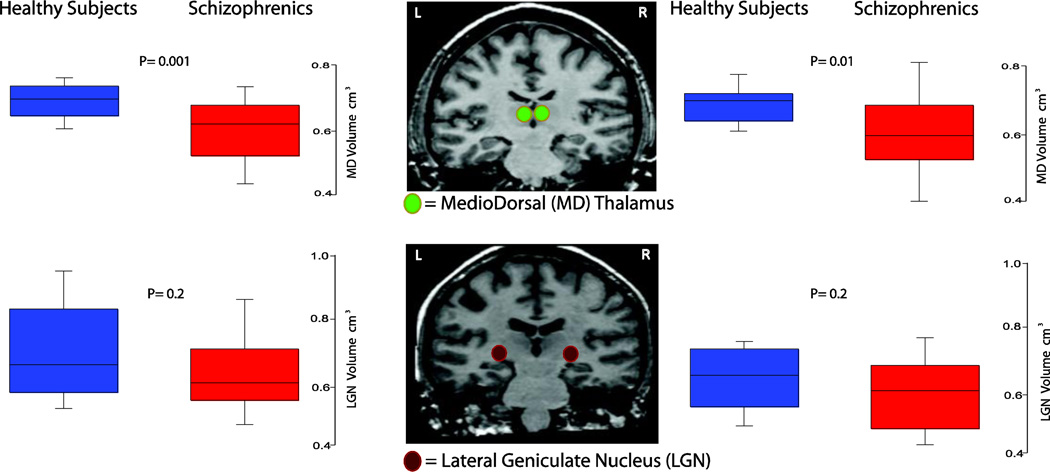
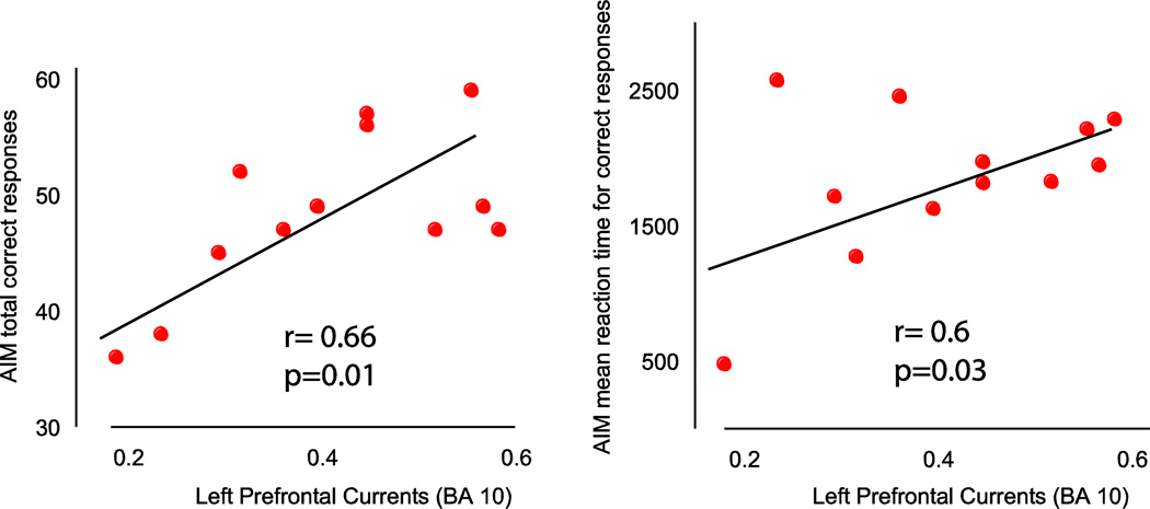
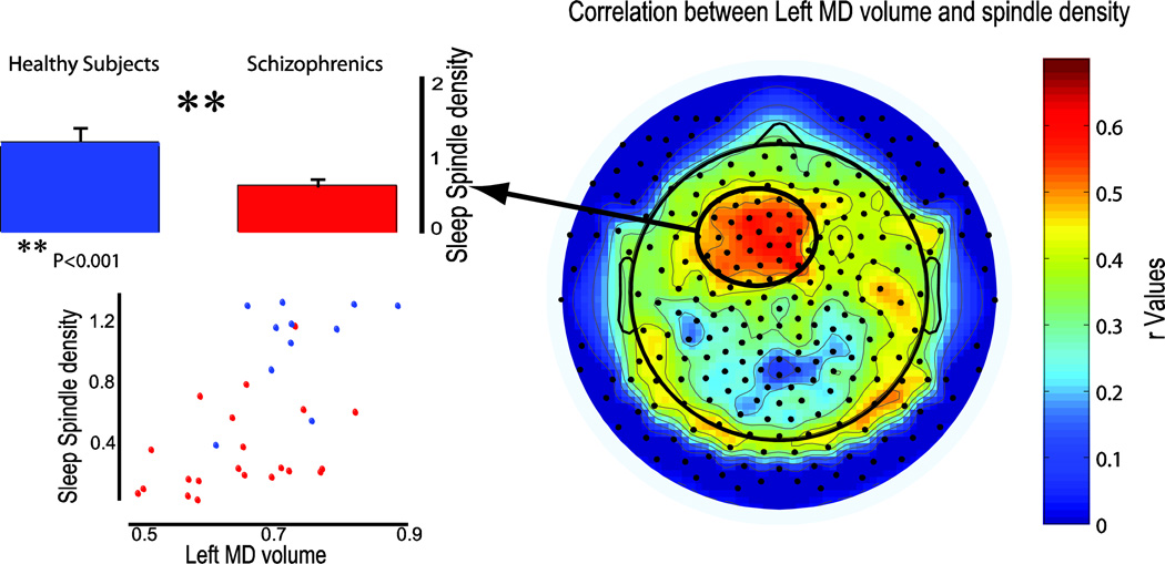
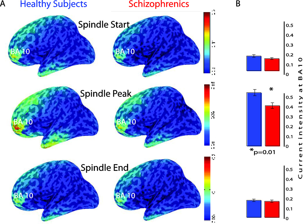
Schizophrenia patients had reduced mediodorsal (MD) thalamic volumes, especially on the left side, compared to healthy controls, whereas whole thalami and lateral geniculate nuclei did not differ between groups. Furthermore, left MD volumes were strongly correlated with the number of scalp-recorded spindles in an anterior frontal region, and cortical currents underlying these anterior frontal spindles were localized in the prefrontal cortex, in Brodmann area (BA) 10. Finally, prefrontal currents at the peak of spindle activity were significantly reduced in schizophrenia patients and correlated with their performance in an abstraction/working memory task.
Altogether, these findings point to deficits in a specific thalamo-cortical circuitry in schizophrenia, which is associated with some cognitive deficits commonly reported in those patients.
我们最近发现,与健康对照者和精神疾病对照者相比,精神分裂症患者的睡眠纺锤波存在明显缺陷。睡眠纺锤波是非快速眼动(NREM)睡眠期的脑电振荡,由丘脑产生,然后在皮质中放大并持续存在。在此,我们研究了丘脑和皮质对这些睡眠纺锤波缺陷的影响。
对精神分裂症患者和健康对照者进行了感兴趣区解剖学体积分析(即丘脑体积)和脑电图(EEG)源模型分析(即与纺锤波相关的皮质电流)。
与健康对照者相比,精神分裂症患者的丘脑内侧背核(MD)体积减小,尤其是左侧,而两组间的整个丘脑和外侧膝状体核无差异。此外,左侧MD体积与前额叶前部头皮记录的纺锤波数量密切相关,这些前额叶前部纺锤波的皮质电流定位于前额叶皮质的Brodmann区(BA)10。最后,精神分裂症患者在纺锤波活动峰值时的前额叶电流显著降低,且与他们在抽象/工作记忆任务中的表现相关。
总之,这些发现表明精神分裂症患者特定丘脑-皮质神经回路存在缺陷,这与这些患者常见的一些认知缺陷有关。