Division of Pulmonary Critical Care & Sleep Medicine, Department of Medicine, Beth Israel Deaconess Medical Center, Boston, MA, USA.
McCance Center for Brain Health, Massachusetts General Hospital, Boston, MA, USA.
Sleep. 2024 Feb 8;47(2). doi: 10.1093/sleep/zsad294.
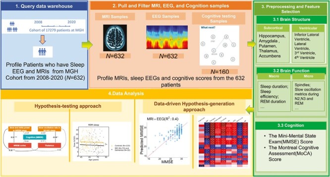
To use relatively noisy routinely collected clinical data (brain magnetic resonance imaging (MRI) data, clinical polysomnography (PSG) recordings, and neuropsychological testing), to investigate hypothesis-driven and data-driven relationships between brain physiology, structure, and cognition.
We analyzed data from patients with clinical PSG, brain MRI, and neuropsychological evaluations. SynthSeg, a neural network-based tool, provided high-quality segmentations despite noise. A priori hypotheses explored associations between brain function (measured by PSG) and brain structure (measured by MRI). Associations with cognitive scores and dementia status were studied. An exploratory data-driven approach investigated age-structure-physiology-cognition links.
Six hundred and twenty-three patients with sleep PSG and brain MRI data were included in this study; 160 with cognitive evaluations. Three hundred and forty-two participants (55%) were female, and age interquartile range was 52 to 69 years. Thirty-six individuals were diagnosed with dementia, 71 with mild cognitive impairment, and 326 with major depression. One hundred and fifteen individuals were evaluated for insomnia and 138 participants had an apnea-hypopnea index equal to or greater than 15. Total PSG delta power correlated positively with frontal lobe/thalamic volumes, and sleep spindle density with thalamic volume. rapid eye movement (REM) duration and amygdala volume were positively associated with cognition. Patients with dementia showed significant differences in five brain structure volumes. REM duration, spindle, and slow-oscillation features had strong associations with cognition and brain structure volumes. PSG and MRI features in combination predicted chronological age (R2 = 0.67) and cognition (R2 = 0.40).
Routine clinical data holds extended value in understanding and even clinically using brain-sleep-cognition relationships.
利用相对嘈杂的常规临床数据(脑磁共振成像(MRI)数据、临床多导睡眠图(PSG)记录和神经心理学测试),研究脑生理学、结构和认知之间的假设驱动和数据驱动关系。
我们分析了来自有临床 PSG、脑 MRI 和神经心理学评估的患者的数据。SynthSeg 是一种基于神经网络的工具,即使在存在噪声的情况下,也能提供高质量的分割。先验假设探讨了脑功能(通过 PSG 测量)与脑结构(通过 MRI 测量)之间的关联。还研究了与认知评分和痴呆状态的关联。探索性的数据驱动方法研究了年龄结构-生理学-认知之间的联系。
本研究纳入了 623 名有睡眠 PSG 和脑 MRI 数据的患者,其中 160 名有认知评估。342 名参与者(55%)为女性,年龄的四分位间距为 52 至 69 岁。36 人被诊断为痴呆症,71 人为轻度认知障碍,326 人为重度抑郁症。115 人接受了失眠评估,138 名参与者的呼吸暂停-低通气指数等于或大于 15。总 PSG 德尔塔功率与额叶/丘脑体积呈正相关,而睡眠纺锤波密度与丘脑体积呈正相关。快速眼动(REM)持续时间和杏仁核体积与认知呈正相关。痴呆症患者的五个脑结构体积存在显著差异。REM 持续时间、纺锤波和慢波振荡特征与认知和脑结构体积有很强的关联。PSG 和 MRI 特征的结合可以预测年龄(R2=0.67)和认知(R2=0.40)。
常规临床数据在理解甚至在临床中使用脑-睡眠-认知关系方面具有扩展价值。