• 文献检索
  • 文档翻译
  • 深度研究
  • 学术资讯
  • Suppr Zotero 插件Zotero 插件
  • 邀请有礼
  • 套餐&价格
  • 历史记录
应用&插件
Suppr Zotero 插件Zotero 插件浏览器插件Mac 客户端Windows 客户端微信小程序
定价
高级版会员购买积分包购买API积分包
服务
文献检索文档翻译深度研究API 文档MCP 服务
关于我们
关于 Suppr公司介绍联系我们用户协议隐私条款
关注我们

Suppr 超能文献

核心技术专利:CN118964589B侵权必究
粤ICP备2023148730 号-1Suppr @ 2026

文献检索

告别复杂PubMed语法,用中文像聊天一样搜索,搜遍4000万医学文献。AI智能推荐,让科研检索更轻松。

立即免费搜索

文件翻译

保留排版,准确专业,支持PDF/Word/PPT等文件格式,支持 12+语言互译。

免费翻译文档

深度研究

AI帮你快速写综述,25分钟生成高质量综述,智能提取关键信息,辅助科研写作。

立即免费体验

双源心脏计算机断层扫描中触发类型、管电压和心率对钙化斑块成像的影响:体模研究

Influence of trigger type, tube voltage and heart rate on calcified plaque imaging in dual source cardiac computed tomography: phantom study.

作者信息

Penzkofer Tobias, Donandt Eva, Isfort Peter, Allmendinger Thomas, Kuhl Christiane K, Mahnken Andreas H, Bruners Philipp

机构信息

Department of Diagnostic and Interventional Radiology, Aachen University Hospital, RWTH Aachen University, Pauwelsstr, 30, 52074 Aachen, Germany.

出版信息

BMC Med Imaging. 2014 Sep 1;14:30. doi: 10.1186/1471-2342-14-30.

DOI:10.1186/1471-2342-14-30
PMID:25178653
原文链接:https://pmc.ncbi.nlm.nih.gov/articles/PMC4174388/
Abstract

BACKGROUND

To investigate the impact of high pitch cardiac CT vs. retrospective ECG gated CT on the quantification of calcified vessel stenoses, with assessment of the influence of tube voltage, reconstruction kernel and heart rate.

METHODS

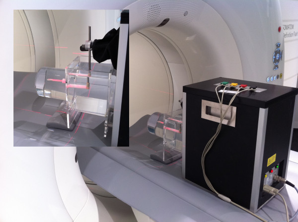
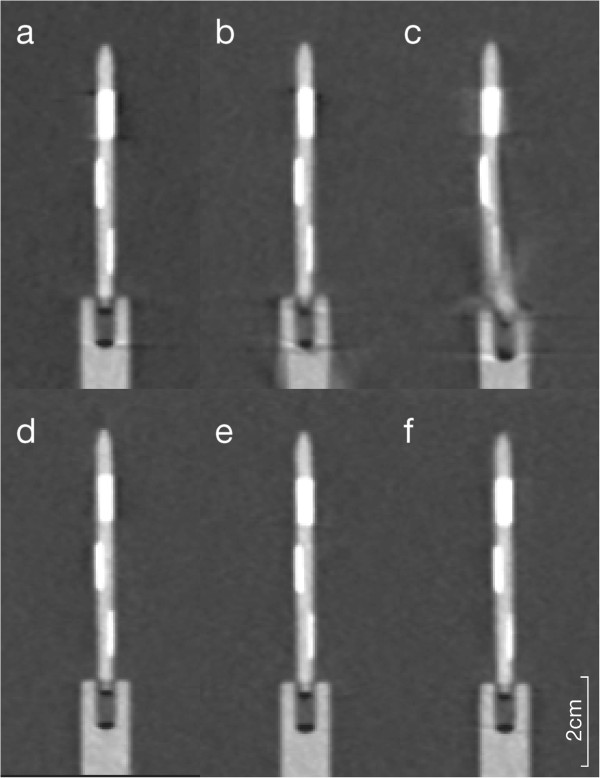
A 4D cardiac movement phantom equipped with three different plaque phantoms (12.5%, 25% and 50% stenosis at different calcification levels), was scanned with a 128-row dual source CT scanner, applying different trigger types (gated vs. prospectively triggered high pitch), tube voltages (100-120 kV) and heart rates (50-90 beats per minute, bpm). Images were reconstructed using different standard (B26f, B46f, B70f) and iterative (I26f, I70f) convolution kernels. Absolute and relative plaque sizes were measured and statistically compared. Radiation dose associated with the different methods (gated vs. high pitch, 100 kV vs. 120 kV) were compared.

RESULTS

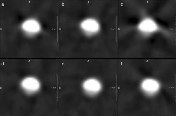
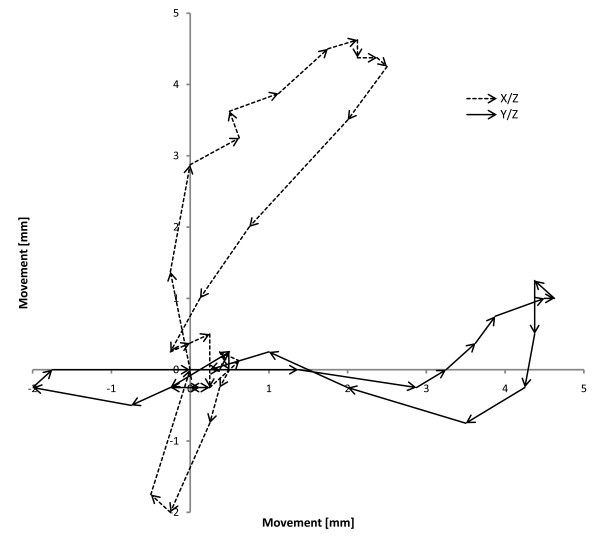
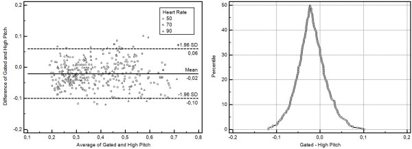
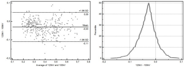
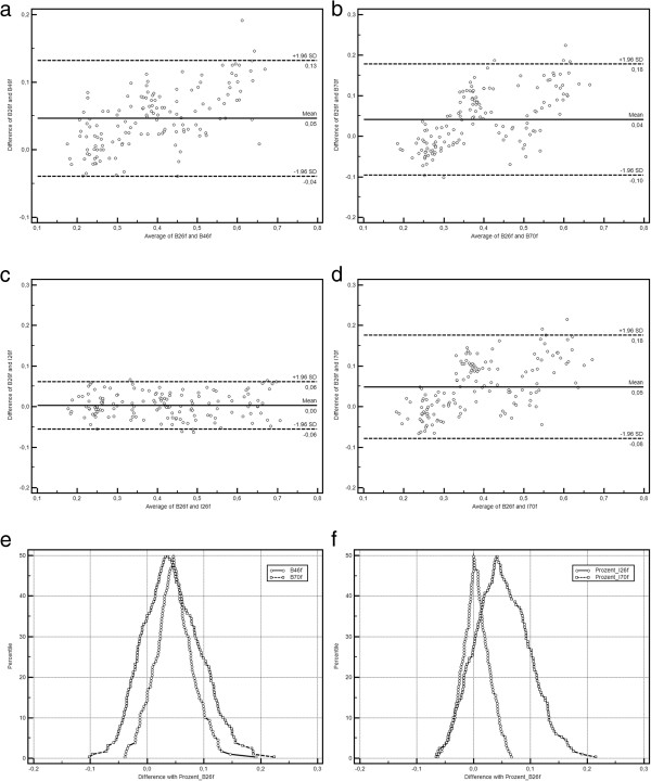
Compared to the known diameters of the phantom plaques and vessels both CT-examination techniques overestimated the degrees of stenoses. Using the high pitch CT-protocol plaques appeared larger (0.09 ± 0.31 mm, 2 ± 8 percent points, PP) in comparison to the ECG-gated CT-scans. Reducing tube voltage had a similar effect, resulting in higher grading of the same stenoses by 3 ± 8 PP. In turn, sharper convolution kernels lead to a lower grading of stenoses (differences of up to 5%). Pairwise comparison of B26f and I26f, B46f and B70f, and B70f and I70f showed differences of 0-1 ± 6-8 PP of the plaque depiction. Motion artifacts were present only at 90 bpm high pitch experiments. High-pitch protocols were associated with significantly lower radiation doses compared with the ECG-gated protocols (258.0 mGy vs. 2829.8 mGy CTDIvol, p ≤ 0.0001).

CONCLUSION

Prospectively triggered high-pitch cardiac CT led to an overestimation of plaque diameter and degree of stenoses in a coronary phantom. This overestimation is only slight and probably negligible in a clinical situation. Even at higher heart rates high pitch CT-scanning allowed reliable measurements of plaque and vessel diameters with only slight differences compared ECG-gated protocols, although motion artifacts were present at 90 bpm using the high pitch protocols.

摘要

背景

研究高螺距心脏CT与回顾性心电图门控CT对钙化血管狭窄定量的影响,并评估管电压、重建核和心率的影响。

方法

使用配备三种不同斑块模型(不同钙化水平下狭窄率分别为12.5%、25%和50%)的4D心脏运动模型,通过128排双源CT扫描仪进行扫描,应用不同的触发类型(门控与前瞻性触发高螺距)、管电压(100 - 120 kV)和心率(每分钟50 - 90次心跳,bpm)。使用不同的标准(B26f、B46f、B70f)和迭代(I26f、I70f)卷积核重建图像。测量并统计比较绝对和相对斑块大小。比较不同方法(门控与高螺距,100 kV与120 kV)相关的辐射剂量。

结果

与模型斑块和血管的已知直径相比,两种CT检查技术均高估了狭窄程度。与心电图门控CT扫描相比,使用高螺距CT方案时斑块显得更大(0.09 ± 0.31 mm,2 ± 8个百分点,PP)。降低管电压有类似效果,导致相同狭窄的分级更高3 ± 8 PP。反过来,更锐利的卷积核导致狭窄分级更低(差异高达5%)。B26f与I26f、B46f与B70f以及B70f与I70f的两两比较显示斑块描绘差异为0 - 1 ± 6 - 8 PP。仅在90 bpm高螺距实验中出现运动伪影。与心电图门控方案相比,高螺距方案的辐射剂量显著更低(容积CT剂量指数CTDIvol为258.0 mGy对2829.8 mGy,p ≤ 0.0001)。

结论

前瞻性触发的高螺距心脏CT导致冠状动脉模型中斑块直径和狭窄程度的高估。这种高估在临床情况下仅轻微且可能可忽略不计。即使在较高心率下,高螺距CT扫描也能可靠测量斑块和血管直径,与心电图门控方案相比差异仅轻微,尽管使用高螺距方案在90 bpm时存在运动伪影。

https://cdn.ncbi.nlm.nih.gov/pmc/blobs/7333/4174388/46a49f8443a5/1471-2342-14-30-8.jpg
https://cdn.ncbi.nlm.nih.gov/pmc/blobs/7333/4174388/4ee94f3a6581/1471-2342-14-30-1.jpg
https://cdn.ncbi.nlm.nih.gov/pmc/blobs/7333/4174388/61ceaf00a789/1471-2342-14-30-2.jpg
https://cdn.ncbi.nlm.nih.gov/pmc/blobs/7333/4174388/3d506e0ee32b/1471-2342-14-30-3.jpg
https://cdn.ncbi.nlm.nih.gov/pmc/blobs/7333/4174388/3b478ba6d69d/1471-2342-14-30-4.jpg
https://cdn.ncbi.nlm.nih.gov/pmc/blobs/7333/4174388/39cb8d66743d/1471-2342-14-30-5.jpg
https://cdn.ncbi.nlm.nih.gov/pmc/blobs/7333/4174388/47153ac564f6/1471-2342-14-30-6.jpg
https://cdn.ncbi.nlm.nih.gov/pmc/blobs/7333/4174388/f2939dd73637/1471-2342-14-30-7.jpg
https://cdn.ncbi.nlm.nih.gov/pmc/blobs/7333/4174388/46a49f8443a5/1471-2342-14-30-8.jpg

相似文献

1
Influence of trigger type, tube voltage and heart rate on calcified plaque imaging in dual source cardiac computed tomography: phantom study.双源心脏计算机断层扫描中触发类型、管电压和心率对钙化斑块成像的影响:体模研究
BMC Med Imaging. 2014 Sep 1;14:30. doi: 10.1186/1471-2342-14-30.
2
The effect of heart rate, vessel angulation and acquisition protocol on the estimation accuracy of calcified artery stenosis in dual energy cardiac CT: A phantom study.心率、血管成角和采集方案对双能心脏 CT 计算钙化动脉狭窄程度准确性的影响:一项体模研究。
Phys Med. 2020 Feb;70:208-215. doi: 10.1016/j.ejmp.2020.02.004. Epub 2020 Feb 12.
3
Very low-dose coronary artery calcium scanning with high-pitch spiral acquisition mode: comparison between 120-kV and 100-kV tube voltage protocols.采用高螺距螺旋采集模式进行超低剂量冠状动脉钙化扫描:120kV 和 100kV 管电压方案的比较。
J Cardiovasc Comput Tomogr. 2013 Jan-Feb;7(1):32-8. doi: 10.1016/j.jcct.2012.11.004. Epub 2012 Dec 1.
4
Radiation dose, image quality, stenosis measurement, and CT densitometry using ECG-triggered coronary 64-MDCT angiography: a phantom study.使用心电图触发的冠状动脉64层螺旋CT血管造影术的辐射剂量、图像质量、狭窄测量及CT密度测定:一项体模研究
AJR Am J Roentgenol. 2008 Feb;190(2):315-20. doi: 10.2214/AJR.07.2191.
5
ECG-triggered high-pitch CT for simultaneous assessment of the aorta and coronary arteries.心电图触发的高分辨率CT用于同时评估主动脉和冠状动脉。
J Cardiovasc Comput Tomogr. 2016 Sep-Oct;10(5):407-13. doi: 10.1016/j.jcct.2016.07.010. Epub 2016 Jul 12.
6
Prospectively ECG-triggered high-pitch coronary angiography with third-generation dual-source CT at 70 kVp tube voltage: feasibility, image quality, radiation dose, and effect of iterative reconstruction.前瞻性心电图触发的70 kVp管电压第三代双源CT高螺距冠状动脉造影:可行性、图像质量、辐射剂量及迭代重建的效果
J Cardiovasc Comput Tomogr. 2014 Nov-Dec;8(6):418-25. doi: 10.1016/j.jcct.2014.09.003. Epub 2014 Sep 16.
7
Dual-source spiral CT with pitch up to 3.2 and 75 ms temporal resolution: image reconstruction and assessment of image quality.采用最大螺距 3.2 和 75 毫秒时间分辨率的双源螺旋 CT:图像重建和图像质量评估。
Med Phys. 2009 Dec;36(12):5641-53. doi: 10.1118/1.3259739.
8
Ultra-high resolution photon-counting coronary CT angiography improves coronary stenosis quantification over a wide range of heart rates - A dynamic phantom study.超高分辨率光子计数冠状动脉 CT 血管造影在较宽心率范围内提高冠状动脉狭窄程度定量评估的准确性:一项动态体模研究。
Eur J Radiol. 2023 Apr;161:110746. doi: 10.1016/j.ejrad.2023.110746. Epub 2023 Feb 16.
9
Systematic analysis on the relationship between luminal enhancement, convolution kernel, plaque density, and luminal diameter of coronary artery stenosis: a CT phantom study.冠状动脉狭窄管腔增强、卷积核、斑块密度与管腔直径关系的系统分析:CT 体模研究。
Int J Cardiovasc Imaging. 2013 Jun;29(5):1129-35. doi: 10.1007/s10554-012-0173-7. Epub 2013 Jan 18.
10
Detection of coronary artery stenoses by low-dose, prospectively ECG-triggered, high-pitch spiral coronary CT angiography.低剂量前瞻性心电图触发高心率螺旋冠状动脉 CT 血管造影检测冠状动脉狭窄。
JACC Cardiovasc Imaging. 2011 Apr;4(4):328-37. doi: 10.1016/j.jcmg.2011.01.012.

引用本文的文献

1
Quantification of uncertainty in the assessment of coronary plaque in CCTA through a dynamic cardiac phantom and 3D-printed plaque model.通过动态心脏模型和3D打印斑块模型对CCTA中冠状动脉斑块评估的不确定性进行量化。
J Med Imaging (Bellingham). 2018 Jan;5(1):013501. doi: 10.1117/1.JMI.5.1.013501. Epub 2018 Jan 17.
2
The Diagnostic Performance of Coronary CT Angiography for the Assessment of Coronary Stenosis in Calcified Plaque.冠状动脉CT血管造影对钙化斑块中冠状动脉狭窄评估的诊断性能
PLoS One. 2016 May 5;11(5):e0154852. doi: 10.1371/journal.pone.0154852. eCollection 2016.

本文引用的文献

1
Performance of turbo high-pitch dual-source CT for coronary CT angiography: first ex vivo and patient experience.涡轮高螺距双源CT在冠状动脉CT血管造影中的性能:首次离体和患者体验。
Eur Radiol. 2014 Aug;24(8):1889-95. doi: 10.1007/s00330-014-3209-7. Epub 2014 May 17.
2
High-pitch coronary CT angiography with third generation dual-source CT: limits of heart rate.第三代双源CT的高心率冠状动脉CT血管造影:心率限制
Int J Cardiovasc Imaging. 2014 Aug;30(6):1173-9. doi: 10.1007/s10554-014-0445-5. Epub 2014 May 11.
3
ACR Appropriateness Criteria(®) acute nonspecific chest pain-low probability of coronary artery disease.
ACR 适宜性标准(®):急性非特异性胸痛-冠状动脉疾病低度可能性。
J Am Coll Radiol. 2012 Oct;9(10):745-50. doi: 10.1016/j.jacr.2012.06.032.
4
Minimizing radiation dose for coronary CT angiography.冠状动脉 CT 血管造影的辐射剂量最小化。
Cardiol Clin. 2012 Feb;30(1):9-17. doi: 10.1016/j.ccl.2011.11.003. Epub 2011 Dec 2.
5
Prospectively ECG-triggered high-pitch spiral acquisition for cardiac CT angiography in routine clinical practice: initial results.前瞻性心电图触发高螺距螺旋采集在常规临床实践中的心脏 CT 血管造影:初步结果。
J Thorac Imaging. 2012 May;27(3):194-201. doi: 10.1097/RTI.0b013e318228223f.
6
High-pitch thoracic CT with simultaneous assessment of coronary arteries: effect of heart rate and heart rate variability on image quality and diagnostic accuracy.高分辨率胸部 CT 同时评估冠状动脉:心率和心率变异性对图像质量和诊断准确性的影响。
JACC Cardiovasc Imaging. 2011 Jun;4(6):602-9. doi: 10.1016/j.jcmg.2011.02.014.
7
Detection of coronary artery stenoses by low-dose, prospectively ECG-triggered, high-pitch spiral coronary CT angiography.低剂量前瞻性心电图触发高心率螺旋冠状动脉 CT 血管造影检测冠状动脉狭窄。
JACC Cardiovasc Imaging. 2011 Apr;4(4):328-37. doi: 10.1016/j.jcmg.2011.01.012.
8
Dual-step prospective ECG-triggered 128-slice dual-source CT for evaluation of coronary arteries and cardiac function without heart rate control: a technical note.双步骤前瞻性心电图触发 128 层双源 CT 用于评估冠状动脉和心功能,无需心率控制:技术说明。
Eur Radiol. 2010 Sep;20(9):2092-9. doi: 10.1007/s00330-010-1794-7. Epub 2010 Apr 21.
9
Dual-source spiral CT with pitch up to 3.2 and 75 ms temporal resolution: image reconstruction and assessment of image quality.采用最大螺距 3.2 和 75 毫秒时间分辨率的双源螺旋 CT:图像重建和图像质量评估。
Med Phys. 2009 Dec;36(12):5641-53. doi: 10.1118/1.3259739.
10
Coronary computed tomography angiography with a consistent dose below 1 mSv using prospectively electrocardiogram-triggered high-pitch spiral acquisition.采用前瞻性心电门控高螺距螺旋扫描技术,使冠状动脉 CT 血管造影的剂量稳定在 1 mSv 以下。
Eur Heart J. 2010 Feb;31(3):340-6. doi: 10.1093/eurheartj/ehp470. Epub 2009 Nov 5.