Noonan MaryAnn P, Sallet Jerome, Mars Rogier B, Neubert Franz X, O'Reilly Jill X, Andersson Jesper L, Mitchell Anna S, Bell Andrew H, Miller Karla L, Rushworth Matthew F S
Department of Experimental Psychology, University of Oxford, Oxford, United Kingdom.
Department of Experimental Psychology, University of Oxford, Oxford, United Kingdom; The Oxford Centre for Functional MRI of the Brain, Nuffield Department of Clinical Neurosciences, University of Oxford, Oxford, United Kingdom.
PLoS Biol. 2014 Sep 2;12(9):e1001940. doi: 10.1371/journal.pbio.1001940. eCollection 2014 Sep.
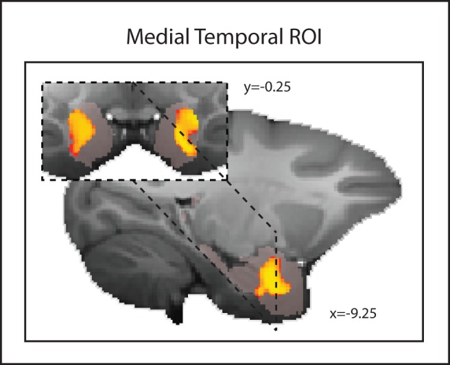
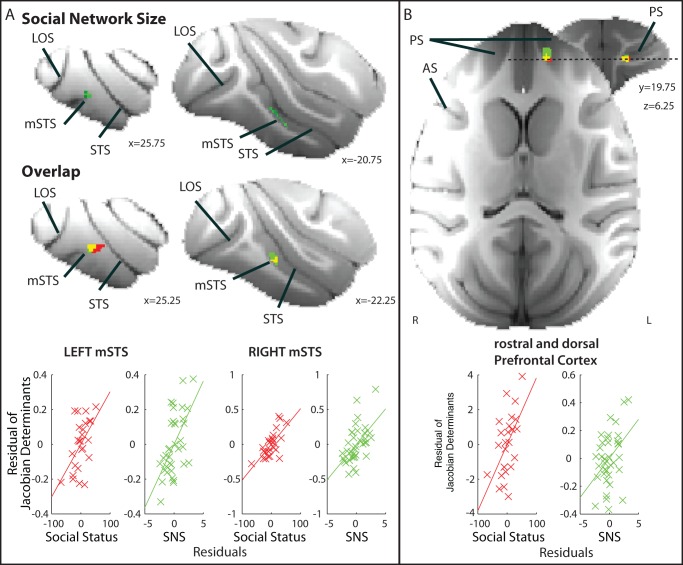
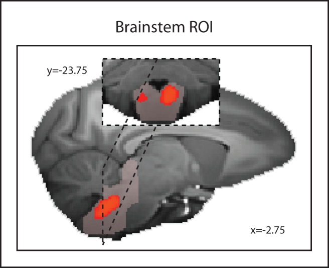
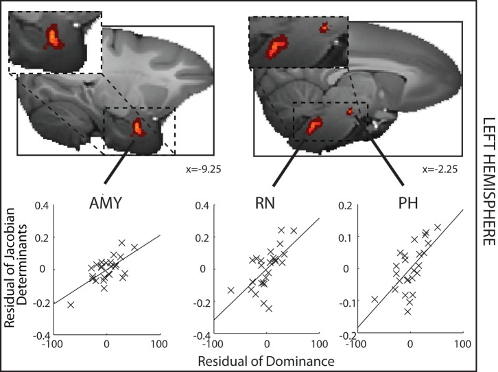
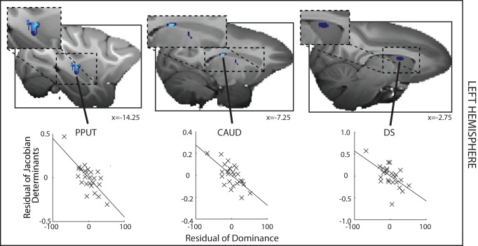
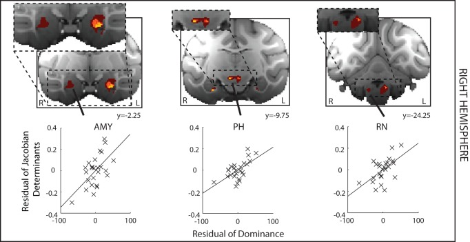
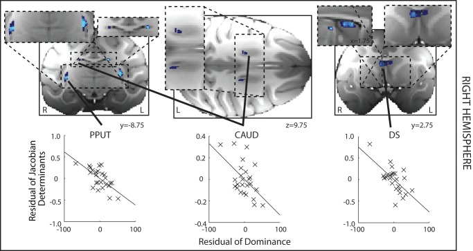
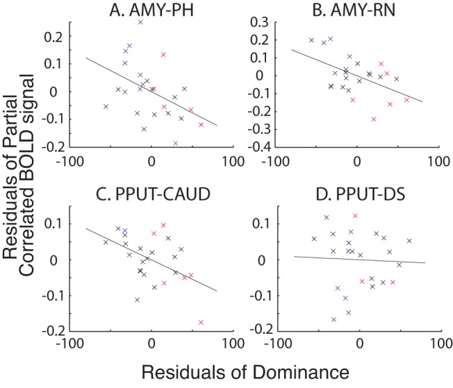
Despite widespread interest in social dominance, little is known of its neural correlates in primates. We hypothesized that social status in primates might be related to individual variation in subcortical brain regions implicated in other aspects of social and emotional behavior in other mammals. To examine this possibility we used magnetic resonance imaging (MRI), which affords the taking of quantitative measurements noninvasively, both of brain structure and of brain function, across many regions simultaneously. We carried out a series of tests of structural and functional MRI (fMRI) data in 25 group-living macaques. First, a deformation-based morphometric (DBM) approach was used to show that gray matter in the amygdala, brainstem in the vicinity of the raphe nucleus, and reticular formation, hypothalamus, and septum/striatum of the left hemisphere was correlated with social status. Second, similar correlations were found in the same areas in the other hemisphere. Third, similar correlations were found in a second data set acquired several months later from a subset of the same animals. Fourth, the strength of coupling between fMRI-measured activity in the same areas was correlated with social status. The network of subcortical areas, however, had no relationship with the sizes of individuals' social networks, suggesting the areas had a simple and direct relationship with social status. By contrast a second circuit in cortex, comprising the midsuperior temporal sulcus and anterior and dorsal prefrontal cortex, covaried with both individuals' social statuses and the social network sizes they experienced. This cortical circuit may be linked to the social cognitive processes that are taxed by life in more complex social networks and that must also be used if an animal is to achieve a high social status.
尽管人们对社会等级制度普遍感兴趣,但对于灵长类动物中其神经关联却知之甚少。我们推测,灵长类动物的社会地位可能与皮质下脑区的个体差异有关,这些脑区在其他哺乳动物的社会和情感行为的其他方面也有涉及。为了检验这种可能性,我们使用了磁共振成像(MRI),它能够同时在多个区域非侵入性地对脑结构和脑功能进行定量测量。我们对25只群居猕猴进行了一系列结构和功能MRI(fMRI)数据测试。首先,基于变形的形态测量(DBM)方法表明,杏仁核中的灰质、中缝核附近的脑干、网状结构、下丘脑以及左半球的隔区/纹状体与社会地位相关。其次,在另一个半球的相同区域也发现了类似的相关性。第三,在几个月后从同一组动物的子集中获取的第二个数据集中也发现了类似的相关性。第四,fMRI测量的相同区域活动之间的耦合强度与社会地位相关。然而,皮质下区域网络与个体社会网络的大小没有关系,这表明这些区域与社会地位有着简单而直接的关系。相比之下,皮质中的第二个回路,包括颞上沟中部以及前额叶背侧和腹侧皮质,与个体的社会地位及其所经历的社会网络大小都存在协变关系。这个皮质回路可能与社会认知过程有关,在更复杂的社会网络中生活时,这些过程会面临挑战,而且如果动物要获得较高的社会地位,也必须运用这些过程。