• 文献检索
  • 文档翻译
  • 深度研究
  • 学术资讯
  • Suppr Zotero 插件Zotero 插件
  • 邀请有礼
  • 套餐&价格
  • 历史记录
应用&插件
Suppr Zotero 插件Zotero 插件浏览器插件Mac 客户端Windows 客户端微信小程序
定价
高级版会员购买积分包购买API积分包
服务
文献检索文档翻译深度研究API 文档MCP 服务
关于我们
关于 Suppr公司介绍联系我们用户协议隐私条款
关注我们

Suppr 超能文献

核心技术专利:CN118964589B侵权必究
粤ICP备2023148730 号-1Suppr @ 2026

文献检索

告别复杂PubMed语法,用中文像聊天一样搜索,搜遍4000万医学文献。AI智能推荐,让科研检索更轻松。

立即免费搜索

文件翻译

保留排版,准确专业,支持PDF/Word/PPT等文件格式,支持 12+语言互译。

免费翻译文档

深度研究

AI帮你快速写综述,25分钟生成高质量综述,智能提取关键信息,辅助科研写作。

立即免费体验

羊膜动物心脏室间隔的演化与发育

Evolution and development of ventricular septation in the amniote heart.

作者信息

Poelmann Robert E, Gittenberger-de Groot Adriana C, Vicente-Steijn Rebecca, Wisse Lambertus J, Bartelings Margot M, Everts Sonja, Hoppenbrouwers Tamara, Kruithof Boudewijn P T, Jensen Bjarke, de Bruin Paul W, Hirasawa Tatsuya, Kuratani Shigeru, Vonk Freek, van de Put Jeanne M M S, de Bakker Merijn A, Richardson Michael K

机构信息

Department of Anatomy and Embryology, Leiden University Medical Center, Leiden, The Netherlands; Department of Cardiology, Leiden University Medical Center, Leiden, The Netherlands; Institute of Biology Leiden (IBL), Leiden University, Sylvius Laboratory, Leiden, The Netherlands.

Department of Cardiology, Leiden University Medical Center, Leiden, The Netherlands.

出版信息

PLoS One. 2014 Sep 5;9(9):e106569. doi: 10.1371/journal.pone.0106569. eCollection 2014.

DOI:10.1371/journal.pone.0106569
PMID:25192012
原文链接:https://pmc.ncbi.nlm.nih.gov/articles/PMC4156344/
Abstract

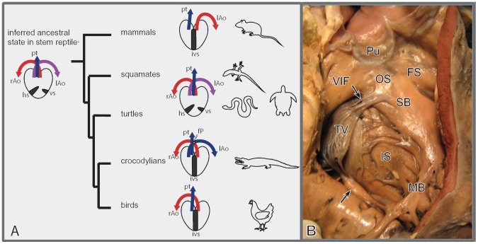
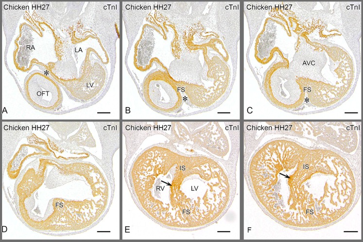
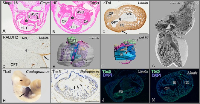
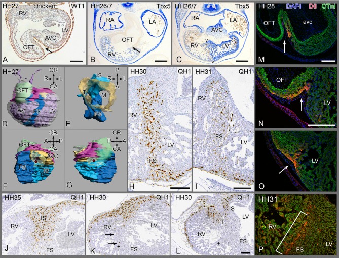
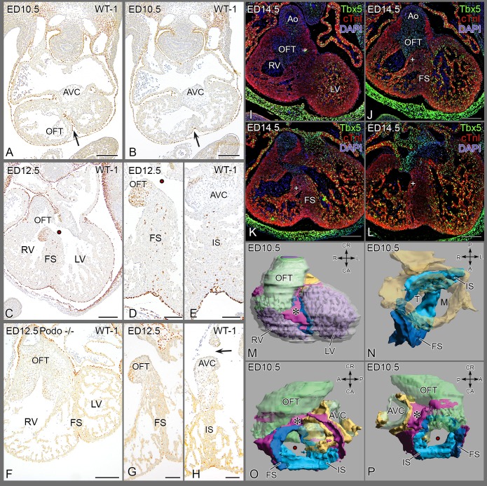
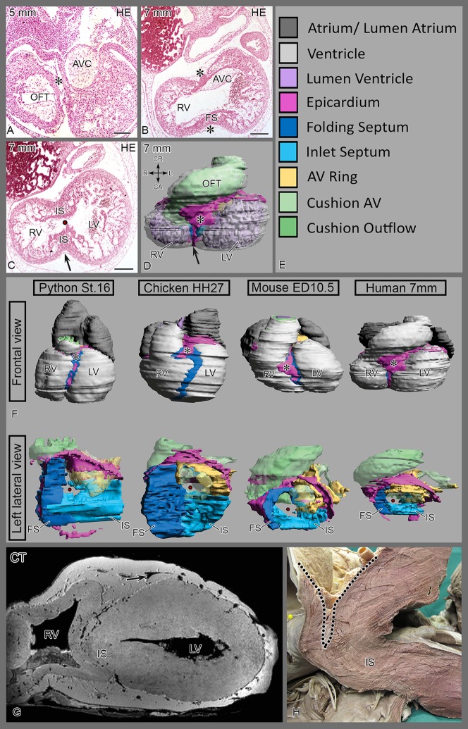
During cardiogenesis the epicardium, covering the surface of the myocardial tube, has been ascribed several functions essential for normal heart development of vertebrates from lampreys to mammals. We investigated a novel function of the epicardium in ventricular development in species with partial and complete septation. These species include reptiles, birds and mammals. Adult turtles, lizards and snakes have a complex ventricle with three cava, partially separated by the horizontal and vertical septa. The crocodilians, birds and mammals with origins some 100 million years apart, however, have a left and right ventricle that are completely separated, being a clear example of convergent evolution. In specific embryonic stages these species show similarities in development, prompting us to investigate the mechanisms underlying epicardial involvement. The primitive ventricle of early embryos becomes septated by folding and fusion of the anterior ventricular wall, trapping epicardium in its core. This folding septum develops as the horizontal septum in reptiles and the anterior part of the interventricular septum in the other taxa. The mechanism of folding is confirmed using DiI tattoos of the ventricular surface. Trapping of epicardium-derived cells is studied by transplanting embryonic quail pro-epicardial organ into chicken hosts. The effect of decreased epicardium involvement is studied in knock-out mice, and pro-epicardium ablated chicken, resulting in diminished and even absent septum formation. Proper folding followed by diminished ventricular fusion may explain the deep interventricular cleft observed in elephants. The vertical septum, although indistinct in most reptiles except in crocodilians and pythonidsis apparently homologous to the inlet septum. Eventually the various septal components merge to form the completely septated heart. In our attempt to discover homologies between the various septum components we aim to elucidate the evolution and development of this part of the vertebrate heart as well as understand the etiology of septal defects in human congenital heart malformations.

摘要

在心脏发生过程中,覆盖心肌管表面的心外膜被认为具有对从七鳃鳗到哺乳动物等脊椎动物正常心脏发育至关重要的多种功能。我们研究了心外膜在部分和完全分隔物种心室发育中的新功能。这些物种包括爬行动物、鸟类和哺乳动物。成年海龟、蜥蜴和蛇有一个复杂的心室,有三个腔,由水平和垂直隔膜部分分隔。然而,起源相隔约1亿年的鳄鱼、鸟类和哺乳动物有完全分隔的左心室和右心室,这是趋同进化的一个明显例子。在特定的胚胎阶段,这些物种在发育上表现出相似性,促使我们研究心外膜参与的潜在机制。早期胚胎的原始心室通过前室壁的折叠和融合而分隔,将心外膜困在其核心。这种折叠隔膜在爬行动物中发育为水平隔膜,在其他分类群中发育为室间隔的前部。通过心室表面的DiI标记证实了折叠机制。通过将胚胎鹌鹑原心外膜器官移植到鸡宿主中来研究心外膜来源细胞的捕获。在基因敲除小鼠和心外膜消融的鸡中研究心外膜参与减少的影响,导致隔膜形成减少甚至缺失。适当的折叠随后心室融合减少可能解释了大象中观察到的深室间裂。垂直隔膜在大多数爬行动物中不明显,除了鳄鱼和蟒科动物,显然与入口隔膜同源。最终,各种隔膜成分合并形成完全分隔的心脏。在我们试图发现各种隔膜成分之间的同源性时,我们旨在阐明脊椎动物心脏这一部分的进化和发育,以及了解人类先天性心脏畸形中隔膜缺损的病因。

https://cdn.ncbi.nlm.nih.gov/pmc/blobs/7079/4156344/2699045dcef7/pone.0106569.g006.jpg
https://cdn.ncbi.nlm.nih.gov/pmc/blobs/7079/4156344/edfb8bfa9fc0/pone.0106569.g001.jpg
https://cdn.ncbi.nlm.nih.gov/pmc/blobs/7079/4156344/232f213e6674/pone.0106569.g002.jpg
https://cdn.ncbi.nlm.nih.gov/pmc/blobs/7079/4156344/69ff488ca6d6/pone.0106569.g003.jpg
https://cdn.ncbi.nlm.nih.gov/pmc/blobs/7079/4156344/fa1c2fdebcc7/pone.0106569.g004.jpg
https://cdn.ncbi.nlm.nih.gov/pmc/blobs/7079/4156344/fabbbe9773b0/pone.0106569.g005.jpg
https://cdn.ncbi.nlm.nih.gov/pmc/blobs/7079/4156344/2699045dcef7/pone.0106569.g006.jpg

相似文献

1
Evolution and development of ventricular septation in the amniote heart.羊膜动物心脏室间隔的演化与发育
PLoS One. 2014 Sep 5;9(9):e106569. doi: 10.1371/journal.pone.0106569. eCollection 2014.
2
The Epicardium in Ventricular Septation During Evolution and Development进化与发育过程中心室分隔中的心外膜
3
Reptilian heart development and the molecular basis of cardiac chamber evolution.爬行动物心脏发育与心腔进化的分子基础。
Nature. 2009 Sep 3;461(7260):95-8. doi: 10.1038/nature08324.
4
Role of the left interventricular sulcus in formation of interventricular septum and crista supraventricularis in normal human cardiogenesis.左室间沟在正常人类心脏发生过程中对室间隔和室上嵴形成的作用。
Anat Rec. 1979 Jul;194(3):417-28. doi: 10.1002/ar.1091940308.
5
New findings concerning ventricular septation in the human heart. Implications for maldevelopment.关于人类心脏室间隔形成的新发现。对发育异常的影响。
Circulation. 1992 Oct;86(4):1194-205. doi: 10.1161/01.cir.86.4.1194.
6
Reptiles as a Model System to Study Heart Development.爬行动物作为研究心脏发育的模型系统。
Cold Spring Harb Perspect Biol. 2020 May 1;12(5):a037226. doi: 10.1101/cshperspect.a037226.
7
Tbx5 specifies the left/right ventricles and ventricular septum position during cardiogenesis.Tbx5在心脏发生过程中确定左/右心室和室间隔的位置。
Development. 2003 Dec;130(24):5953-64. doi: 10.1242/dev.00797. Epub 2003 Oct 22.
8
Altered haemodynamics causes aberrations in the epicardium.改变血流动力学导致心外膜出现异常。
J Anat. 2019 Jun;234(6):800-814. doi: 10.1111/joa.12977. Epub 2019 Mar 18.
9
The septation of the arterial pole of the heart in the chick embryo. III. Development of the truncus arteriosus of the heart of chick embryos from 5 1/2 to 7 days of incubation.鸡胚心脏动脉极的分隔。III. 孵化5.5至7天鸡胚心脏动脉干的发育。
Acta Morphol Neerl Scand. 1979 Feb;17(1):1-20.
10
Outflow tract septation and the aortic arch system in reptiles: lessons for understanding the mammalian heart.爬行动物的流出道分隔与主动脉弓系统:对理解哺乳动物心脏的启示
Evodevo. 2017 May 10;8:9. doi: 10.1186/s13227-017-0072-z. eCollection 2017.

引用本文的文献

1
The Interplay of Ontogeny and Phylogeny at the Transcriptome Level of the Tetrapod Heart.四足动物心脏转录组水平上个体发育与系统发育的相互作用
J Exp Zool B Mol Dev Evol. 2025 Sep;344(6):325-340. doi: 10.1002/jez.b.23312. Epub 2025 Jul 3.
2
A Comprehensive Review of Canine and Feline Ventricular Septal Defects-From Pathogenesis to Long-Term Follow-Up.犬猫室间隔缺损综述——从发病机制到长期随访
Animals (Basel). 2025 Mar 15;15(6):850. doi: 10.3390/ani15060850.
3
The avian embryo as a time-honoured animal model in developmental, biomedical and agricultural research.

本文引用的文献

1
A reconsideration of the phylogeny of the tetrapod heart.对四足动物心脏系统发育的重新思考。
J Morphol. 1975 Oct;147(2):209-228. doi: 10.1002/jmor.1051470207.
2
A series of normal stages in the development of the chick embryo.鸡胚胎发育的一系列正常阶段。
J Morphol. 1951 Jan;88(1):49-92.
3
Structure and function of the hearts of lizards and snakes.蜥蜴和蛇类心脏的结构与功能。
鸟类胚胎作为发育、生物医学和农业研究中历史悠久的动物模型。
Philos Trans R Soc Lond B Biol Sci. 2025 Feb 27;380(1920):20230438. doi: 10.1098/rstb.2023.0438.
4
Ventricular Septal Defects: Molecular Pathways and Animal Models.室间隔缺损:分子途径和动物模型。
Adv Exp Med Biol. 2024;1441:535-549. doi: 10.1007/978-3-031-44087-8_28.
5
Narrative review of single ventricle: where are we after 40 years?单心室的叙述性综述:40年后我们处于什么阶段?
Transl Pediatr. 2023 Feb 28;12(2):221-244. doi: 10.21037/tp-22-573. Epub 2023 Feb 7.
6
Anguimorpha as a model group for studying the comparative heart morphology among Lepidosauria: Evolutionary window on the ventricular septation.有鳞目蜥蜴亚目作为研究比较心脏形态的模型类群:心室间隔的进化窗口。
Ecol Evol. 2022 Nov 8;12(11):e9476. doi: 10.1002/ece3.9476. eCollection 2022 Nov.
7
Single ventricle: amphibians and human beings.单心室:两栖动物与人类。
World J Pediatr. 2022 Oct;18(10):643-646. doi: 10.1007/s12519-022-00595-5.
8
Anatomy of the heart of the leatherback turtle.棱皮龟心脏解剖结构。
J Anat. 2022 Aug;241(2):535-544. doi: 10.1111/joa.13670. Epub 2022 Apr 12.
9
Whole-body endothermy: ancient, homologous and widespread among the ancestors of mammals, birds and crocodylians.全身温血:在哺乳动物、鸟类和鳄类的祖先中,这种现象既古老又同源且广泛存在。
Biol Rev Camb Philos Soc. 2022 Apr;97(2):766-801. doi: 10.1111/brv.12822. Epub 2021 Dec 10.
10
High heart rate associated early repolarization causes J-waves in both zebra finch and mouse.高心率相关的早期复极导致斑马雀和小鼠中 J 波的产生。
Physiol Rep. 2021 Mar;9(5):e14775. doi: 10.14814/phy2.14775.
Biol Rev Camb Philos Soc. 2014 May;89(2):302-36. doi: 10.1111/brv.12056. Epub 2013 Sep 2.
4
Digit loss in archosaur evolution and the interplay between selection and constraints.兽脚亚目恐龙进化中的指节缺失和选择与约束之间的相互作用。
Nature. 2013 Aug 22;500(7463):445-8. doi: 10.1038/nature12336. Epub 2013 Jul 7.
5
Development of the hearts of lizards and snakes and perspectives to cardiac evolution.蜥蜴和蛇类心脏的发育与心脏演化的展望。
PLoS One. 2013 Jun 5;8(6):e63651. doi: 10.1371/journal.pone.0063651. Print 2013.
6
The draft genomes of soft-shell turtle and green sea turtle yield insights into the development and evolution of the turtle-specific body plan.软壳龟和绿海龟的基因组草图为龟类特有的身体结构的发育和进化提供了新的见解。
Nat Genet. 2013 Jun;45(6):701-706. doi: 10.1038/ng.2615. Epub 2013 Apr 28.
7
The Wilms' tumor suppressor Wt1 regulates Coronin 1B expression in the epicardium.Wilms 瘤抑制因子 Wt1 调控心外膜中 Coronin 1B 的表达。
Exp Cell Res. 2013 Jun 10;319(10):1365-81. doi: 10.1016/j.yexcr.2013.03.027. Epub 2013 Apr 4.
8
C/EBP transcription factors mediate epicardial activation during heart development and injury.C/EBP 转录因子在心脏发育和损伤过程中介导心外膜的激活。
Science. 2012 Dec 21;338(6114):1599-603. doi: 10.1126/science.1229765. Epub 2012 Nov 15.
9
Genetic Cre-loxP assessment of epicardial cell fate using Wt1-driven Cre alleles.利用 Wt1 驱动的 Cre 等位基因进行心外膜细胞命运的遗传 Cre-loxP 评估。
Circ Res. 2012 Nov 9;111(11):e276-80. doi: 10.1161/CIRCRESAHA.112.275784.
10
Epicardial progenitor cells in cardiac regeneration and neovascularisation.心外膜祖细胞在心脏再生和血管新生中的作用。
Vascul Pharmacol. 2013 Mar;58(3):164-73. doi: 10.1016/j.vph.2012.08.001. Epub 2012 Aug 11.