Jayasuriya Chathuraka T, Zhou Fiona H, Pei Ming, Wang Zhengke, Lemme Nicholas J, Haines Paul, Chen Qian
Department of Orthopaedics, Warren Alpert Medical School of Brown University, CORO West, Suite 402A, 1 Hoppin Street, Providence, RI 02903, USA.
Stem Cell and Tissue Engineering Laboratory, Department of Orthopaedics, West Virginia University, Morgantown, WV 26506, USA.
Int J Mol Sci. 2014 Aug 21;15(8):14555-73. doi: 10.3390/ijms150814555.
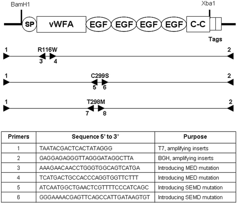
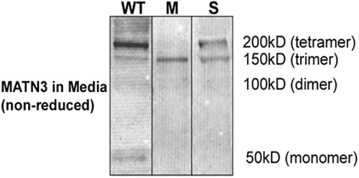
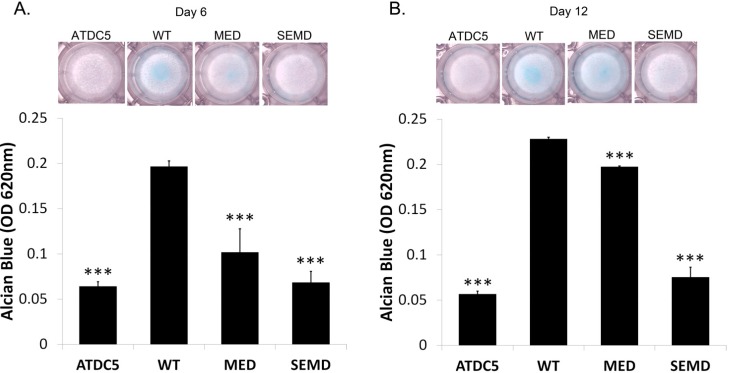
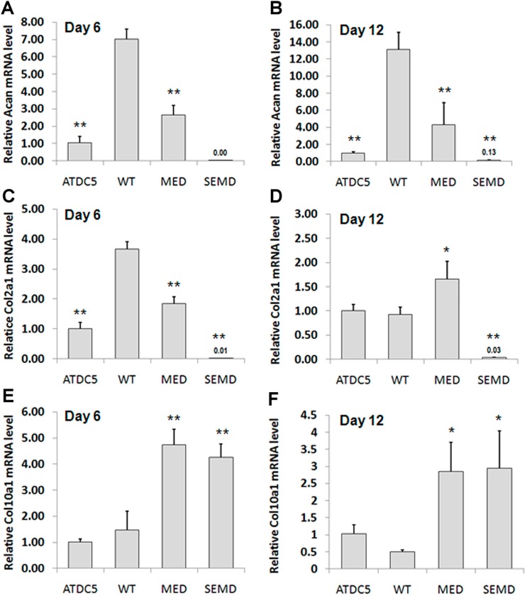
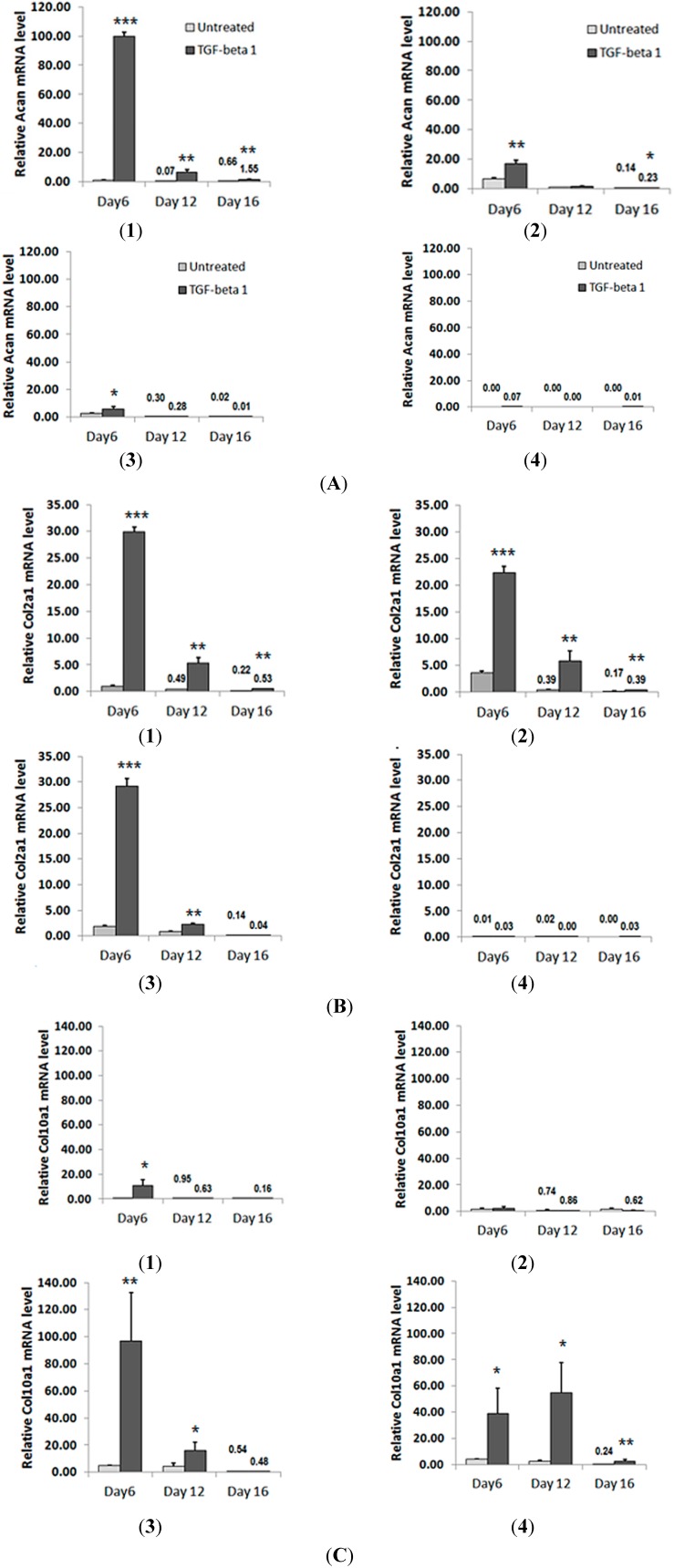
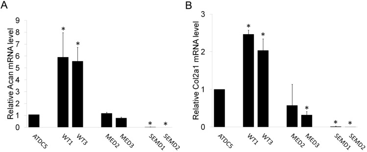
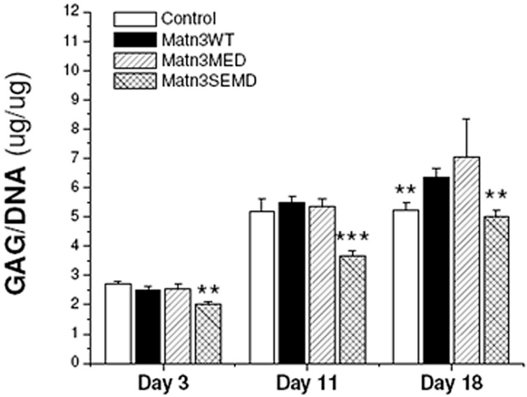
Studies have shown that mutations in the matrilin-3 gene (MATN3) are associated with multiple epiphyseal dysplasia (MED) and spondyloepimetaphyseal dysplasia (SEMD). We tested whether MATN3 mutations affect the differentiation of chondroprogenitor and/or mesenchymal stem cells, which are precursors to chondrocytes. ATDC5 chondroprogenitors stably expressing wild-type (WT) MATN3 underwent spontaneous chondrogenesis. Expression of chondrogenic markers collagen II and aggrecan was inhibited in chondroprogenitors carrying the MED or SEMD MATN3 mutations. Hypertrophic marker collagen X remained attenuated in WT MATN3 chondroprogenitors, whereas its expression was elevated in chondroprogenitors expressing the MED or SEMD mutant MATN3 gene suggesting that these mutations inhibit chondrogenesis but promote hypertrophy. TGF-β treatment failed to rescue chondrogenesis markers but dramatically increased collagen X mRNA expression in mutant MATN3 expressing chondroprogenitors. Synovium derived mesenchymal stem cells harboring the SEMD mutation exhibited lower glycosaminoglycan content than those of WT MATN3 in response to TGF-β. Our results suggest that the properties of progenitor cells harboring MATN3 chondrodysplasia mutations were altered, as evidenced by attenuated chondrogenesis and premature hypertrophy. TGF-β treatment failed to completely rescue chondrogenesis but instead induced hypertrophy in mutant MATN3 chondroprogenitors. Our data suggest that chondroprogenitor cells should be considered as a potential target of chondrodysplasia therapy.
研究表明,基质金属蛋白酶3基因(MATN3)突变与多发性骨骺发育不良(MED)和脊椎骨骺发育不良(SEMD)相关。我们测试了MATN3突变是否会影响软骨祖细胞和/或间充质干细胞(软骨细胞的前体)的分化。稳定表达野生型(WT)MATN3的ATDC5软骨祖细胞发生了自发软骨形成。携带MED或SEMD的MATN3突变的软骨祖细胞中,软骨生成标志物II型胶原蛋白和聚集蛋白聚糖的表达受到抑制。肥大标志物X型胶原蛋白在WT MATN3软骨祖细胞中仍然减弱,而在表达MED或SEMD突变MATN3基因的软骨祖细胞中其表达升高,这表明这些突变抑制软骨生成但促进肥大。TGF-β处理未能挽救软骨生成标志物,但显著增加了表达突变MATN3的软骨祖细胞中X型胶原蛋白mRNA的表达。携带SEMD突变的滑膜来源间充质干细胞在TGF-β刺激下,与WT MATN3相比,糖胺聚糖含量更低。我们的结果表明,携带MATN3软骨发育异常突变的祖细胞特性发生了改变,表现为软骨生成减弱和过早肥大。TGF-β处理未能完全挽救软骨生成,反而在突变MATN3软骨祖细胞中诱导了肥大。我们的数据表明,软骨祖细胞应被视为软骨发育异常治疗的潜在靶点。