Kim Hyonchol, Terazono Hideyuki, Takei Hiroyuki, Yasuda Kenji
Kanagawa Academy of Science and Technology, KSP East 310, 3-2-1 Sakado, Takatsu-ku, Kawasaki, Kanagawa 213-0012, Japan.
1] Kanagawa Academy of Science and Technology, KSP East 310, 3-2-1 Sakado, Takatsu-ku, Kawasaki, Kanagawa 213-0012, Japan [2] Department of Biomedical Information, Division of Biosystems, Institute of Biomaterials and Bioengineering, Tokyo Medical and Dental University, 2-3-10 Kanda-Surugadai, Chiyoda-ku, Tokyo 101-0062, Japan.
Sci Rep. 2014 Sep 15;4:6362. doi: 10.1038/srep06362.
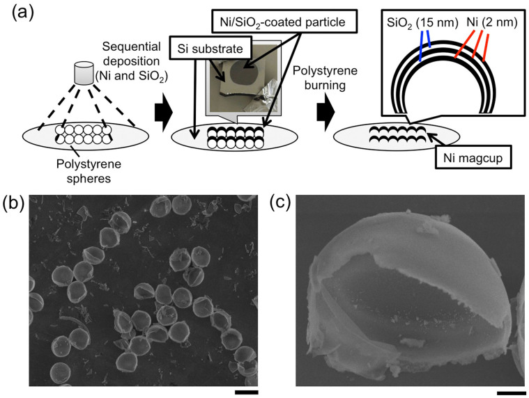
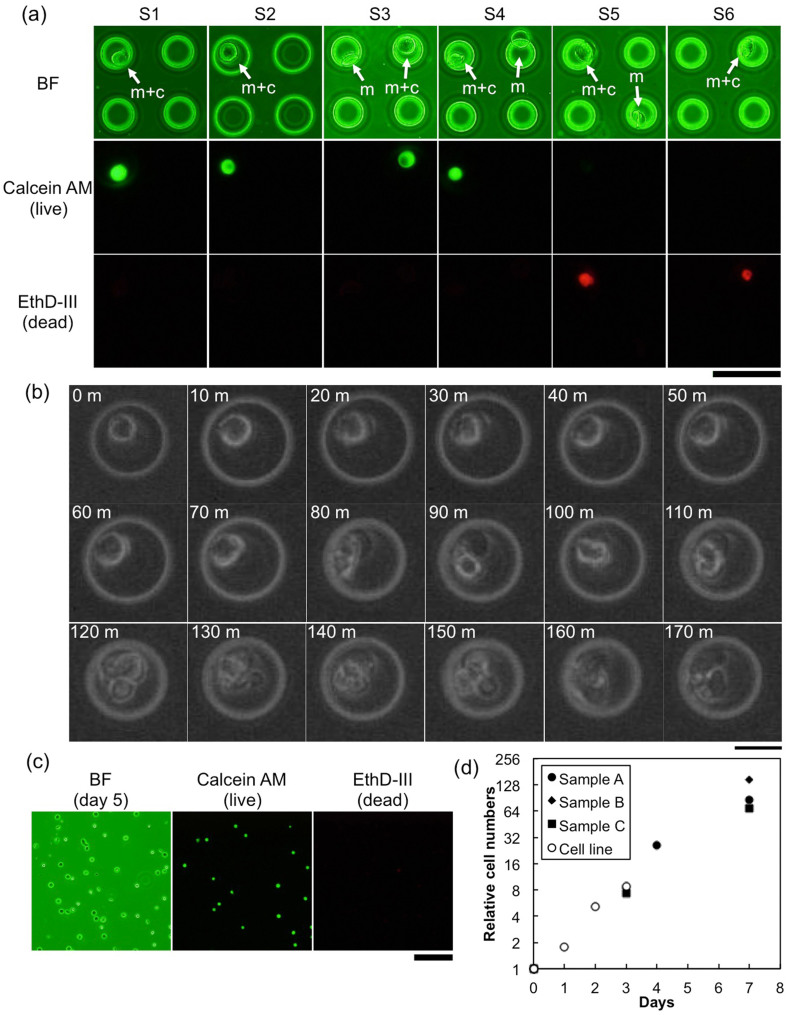
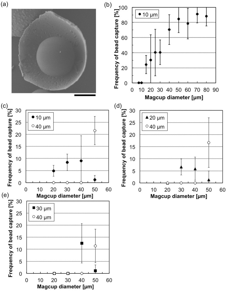
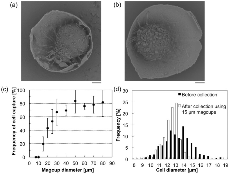
We propose a new method of size separation of cells exploiting precisely size-controlled hemispherical superparamagnetic microparticles. A three-layered structure of a 2-nm nickel layer inserted between 15-nm silicon dioxide layers was formed on polystyrene cast spheres by vapor deposition. The polystyrene was then removed by burning and the hemispherical superparamagnetic microparticles, "magcups", were obtained. The standard target cells (CCRF-CEM, 12 ± 2 μm) were mixed with a set of different sizes of the fabricated magcups, and we confirmed that the cells were captured in the magcups having cavities larger than 15 μm in diameter, and then gathered by magnetic force. The collected cells were grown in a culture medium without any damage. The results suggest that this method is quick, simple and non-invasive size separation of target cells.
我们提出了一种利用精确尺寸控制的半球形超顺磁性微粒对细胞进行尺寸分离的新方法。通过气相沉积在聚苯乙烯铸球上形成了一种三层结构,即在15纳米二氧化硅层之间插入2纳米镍层。然后通过燃烧去除聚苯乙烯,从而获得半球形超顺磁性微粒“磁杯”。将标准靶细胞(CCRF-CEM,12±2μm)与一组不同尺寸的制备好的磁杯混合,我们证实细胞被捕获在直径大于15μm的有腔磁杯中,然后通过磁力聚集。收集到的细胞在培养基中生长且没有任何损伤。结果表明,该方法是一种快速、简单且非侵入性的靶细胞尺寸分离方法。