Cheng Xinlai, Holenya Palvo, Can Suzan, Alborzinia Hamed, Rubbiani Riccardo, Ott Ingo, Wölfl Stefan
Institut für Pharmazie und Molekulare Biotechnologie, Ruprecht-Karls-Universität Heidelberg, Im Neuenheimer Feld 364, 69120 Heidelberg, Germany.
Mol Cancer. 2014 Sep 25;13:221. doi: 10.1186/1476-4598-13-221.
Cancer cells in the advanced stage show aberrant antioxidant capacity to detoxify excessive ROS resulting in the compensation for intrinsic oxidative stress and therapeutic resistance. PDAC is one of the most lethal cancers and often associated with a high accumulation of ROS. Recent studies identified gold(I) NHC complexes as potent TrxR inhibitors suppressing cell growth in a wide spectrum of human malignant cell lines at the low micromolar concentration. However, the mechanism of action is not completely elucidated yet.
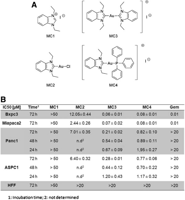
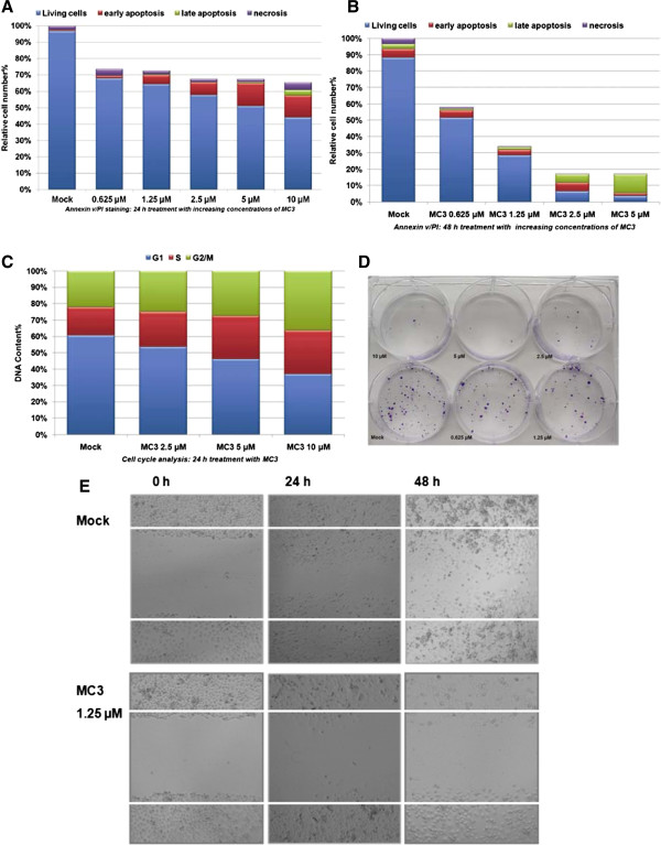
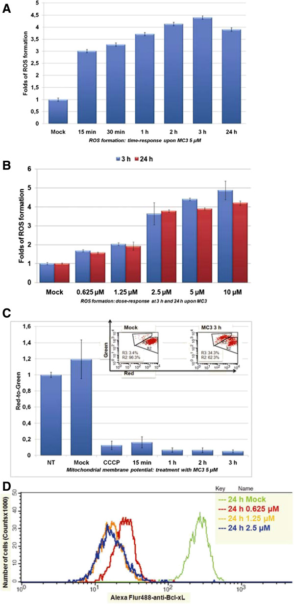
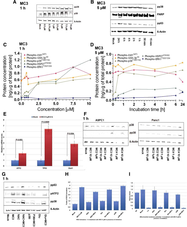
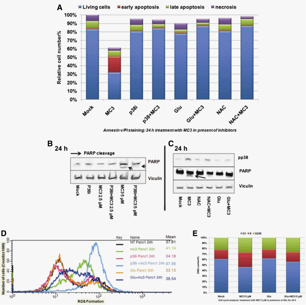
To understand the biological function of gold(I) NHC complexes in PDAC, we used a recently published gold(I) NHC complex, MC3, and evaluated its anti-proliferative effect in four PDAC cell lines, determined by MTT and SRB assays. In further detailed analysis, we analyzed cellular ROS levels using the ROS indicator DHE and mitochondrial membrane potential indicated by the dye JC-1 in Panc1. We also analyzed cell cycle arrest and apoptosis by FACS. To elucidate the role of specific cell signaling pathways in MC3-induced cell death, co-incubation with ROS scavengers, a p38-MAPK inhibitor and siRNA mediated depletion of ASK1 were performed, and results were analyzed by immunoblotting, ELISA-microarrays, qRT-PCR and immunoprecipitation.
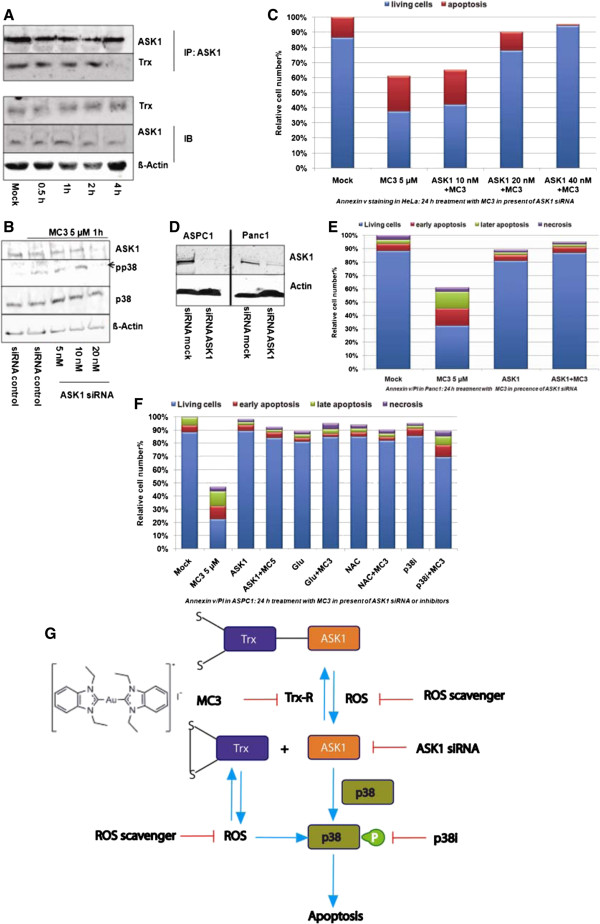
Our data demonstrate that MC3 efficiently suppressed cell growth, and induced cell cycle arrest and apoptosis in pancreatic cancer cells, in particular in the gemcitabine-resistant cancer cells Panc1 and ASPC1. Treatment with MC3 resulted in a substantial alteration of the cellular redox homeostasis leading to increased ROS levels and a decrease in the mitochondrial membrane potential. ROS scavengers suppressed ROS formation and rescued cells from damage. On the molecular level, MC3 blocked the interaction of Trx with ASK1 and subsequently activated p38-associated signaling. Furthermore, inhibition of this pathway by using ASK1 siRNA or a p38 inhibitor clearly attenuated the effect of MC3 on cell proliferation in Panc1 and ASPC1.
Our results confirm that MC3 is a TrxR inhibitor and show MC3 induced apoptosis in gemcitabine-resistant PDACs. MC3 mediated cell death could be blocked by using anti-oxidants, ASK1 siRNA or p38 inhibitor suggesting that the Trx-ASK1-p38 signal cascade played an important role in gold(I) NHC complexes-mediated cellular damage.
晚期癌细胞表现出异常的抗氧化能力,可清除过量的活性氧(ROS),从而补偿内在的氧化应激并产生治疗抗性。胰腺导管腺癌(PDAC)是最致命的癌症之一,常与高水平的ROS积累相关。最近的研究发现金(I)氮杂环卡宾配合物是有效的硫氧还蛋白还原酶(TrxR)抑制剂,能在低微摩尔浓度下抑制多种人类恶性细胞系的细胞生长。然而,其作用机制尚未完全阐明。
为了解金(I)氮杂环卡宾配合物在PDAC中的生物学功能,我们使用了最近发表的金(I)氮杂环卡宾配合物MC3,并通过MTT和SRB试验评估了其在四种PDAC细胞系中的抗增殖作用。在进一步的详细分析中,我们使用ROS指示剂二氢乙锭(DHE)分析了细胞内ROS水平,并使用染料JC-1分析了Panc1细胞中的线粒体膜电位。我们还通过流式细胞术分析了细胞周期阻滞和凋亡。为阐明特定细胞信号通路在MC3诱导的细胞死亡中的作用,我们进行了与ROS清除剂、p38丝裂原活化蛋白激酶(MAPK)抑制剂的共孵育以及小干扰RNA(siRNA)介导的凋亡信号调节激酶1(ASK1)的消耗,并通过免疫印迹、酶联免疫吸附测定微阵列、定量逆转录聚合酶链反应(qRT-PCR)和免疫沉淀分析结果。
我们的数据表明,MC3能有效抑制胰腺癌细胞的生长,并诱导细胞周期阻滞和凋亡,特别是在吉西他滨耐药的癌细胞Panc1和ASPC1中。用MC3处理导致细胞氧化还原稳态发生显著改变,导致ROS水平升高和线粒体膜电位降低。ROS清除剂抑制了ROS的形成并使细胞免受损伤。在分子水平上,MC3阻断了Trx与ASK1的相互作用,随后激活了与p38相关的信号传导。此外,使用ASK1 siRNA或p38抑制剂抑制该信号通路明显减弱了MC3对Panc1和ASPC1细胞增殖的影响。
我们的结果证实MC3是一种TrxR抑制剂,并表明MC3可诱导吉西他滨耐药的PDAC细胞凋亡。使用抗氧化剂、ASK1 siRNA或p38抑制剂可阻断MC3介导的细胞死亡,这表明Trx-ASK1-p38信号级联在金(I)氮杂环卡宾配合物介导的细胞损伤中起重要作用。