Dillon L M, Bean J R, Yang W, Shee K, Symonds L K, Balko J M, McDonald W H, Liu S, Gonzalez-Angulo A M, Mills G B, Arteaga C L, Miller T W
Department of Pharmacology and Toxicology, Norris Cotton Cancer Center, Geisel School of Medicine at Dartmouth, Dartmouth-Hitchcock Medical Center, Lebanon, NH, USA.
1] Department of Medicine, Vanderbilt-Ingram Cancer Center, Vanderbilt University School of Medicine, Nashville, TN, USA [2] Breast Cancer Research Program, Vanderbilt-Ingram Cancer Center, Vanderbilt University School of Medicine, Nashville, TN, USA.
Oncogene. 2015 Jul 23;34(30):3968-76. doi: 10.1038/onc.2014.328. Epub 2014 Oct 6.
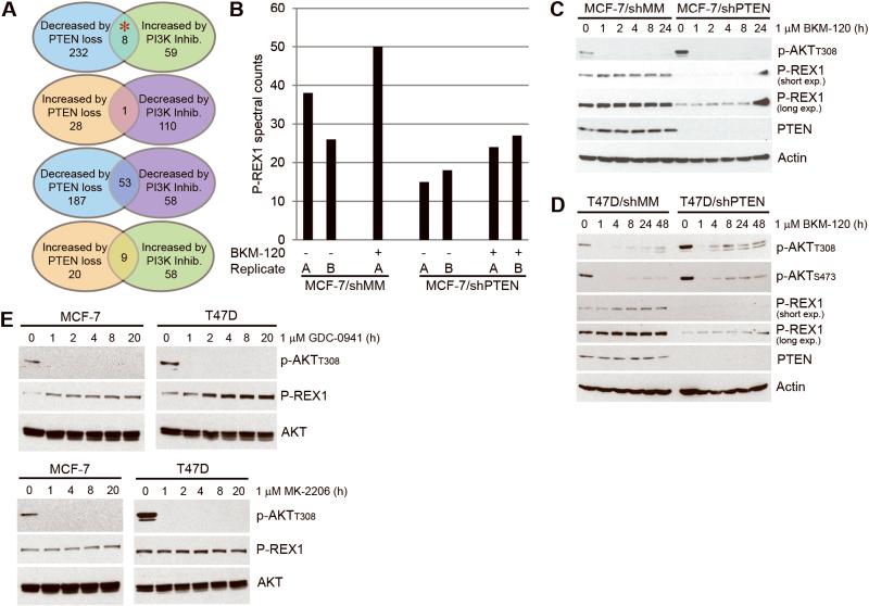
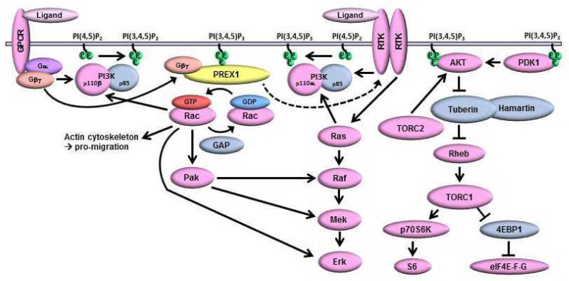
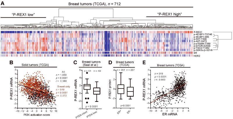
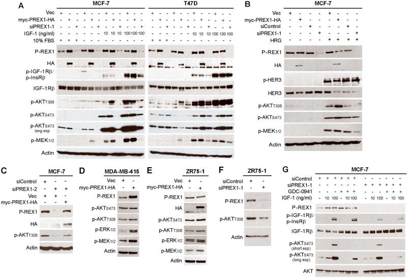
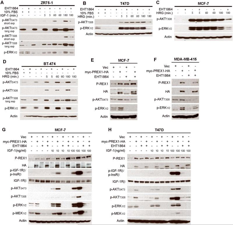
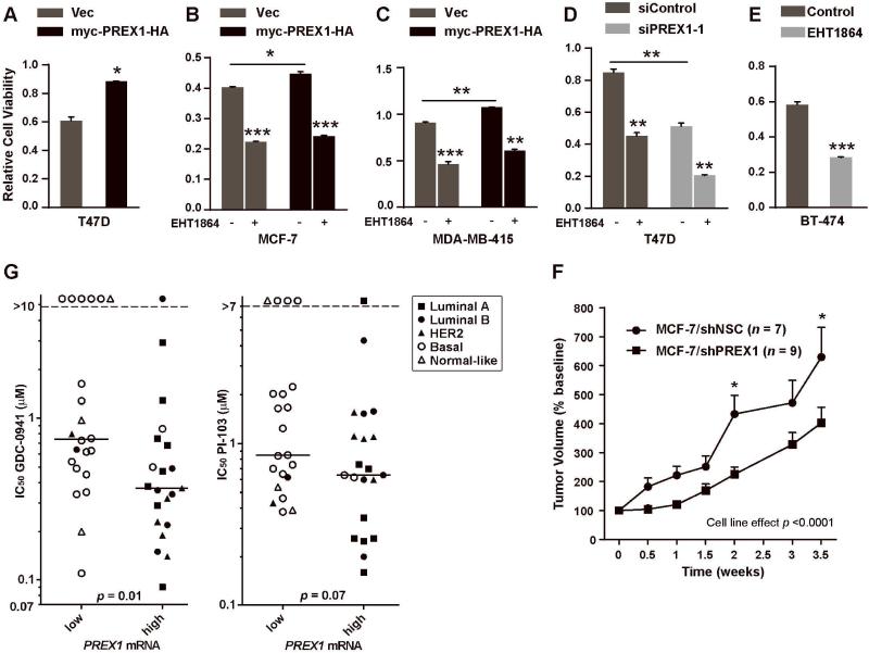
Phosphatidylinositol 3-kinase (PI3K) promotes cancer cell survival, migration, growth and proliferation by generating phosphatidylinositol 3,4,5-trisphosphate (PIP3) in the inner leaflet of the plasma membrane. PIP3 recruits pleckstrin homology domain-containing proteins to the membrane to activate oncogenic signaling cascades. Anticancer therapeutics targeting the PI3K/AKT/mTOR (mammalian target of rapamycin) pathway are in clinical development. In a mass spectrometric screen to identify PIP3-regulated proteins in breast cancer cells, levels of the Rac activator PIP3-dependent Rac exchange factor-1 (P-REX1) increased in response to PI3K inhibition, and decreased upon loss of the PI3K antagonist phosphatase and tensin homolog (PTEN). P-REX1 mRNA and protein levels were positively correlated with ER expression, and inversely correlated with PI3K pathway activation in breast tumors as assessed by gene expression and phosphoproteomic analyses. P-REX1 increased activation of Rac1, PI3K/AKT and MEK/ERK signaling in a PTEN-independent manner, and promoted cell and tumor viability. Loss of P-REX1 or inhibition of Rac suppressed PI3K/AKT and MEK/ERK, and decreased viability. P-REX1 also promoted insulin-like growth factor-1 receptor activation, suggesting that P-REX1 provides positive feedback to activators upstream of PI3K. In support of a model where PIP3-driven P-REX1 promotes both PI3K/AKT and MEK/ERK signaling, high levels of P-REX1 mRNA (but not phospho-AKT or a transcriptomic signature of PI3K activation) were predictive of sensitivity to PI3K inhibitors among breast cancer cell lines. P-REX1 expression was highest in estrogen receptor-positive breast tumors compared with many other cancer subtypes, suggesting that neutralizing the P-REX1/Rac axis may provide a novel therapeutic approach to selectively abrogate oncogenic signaling in breast cancer cells.
磷脂酰肌醇3激酶(PI3K)通过在质膜内小叶中生成磷脂酰肌醇3,4,5-三磷酸(PIP3)来促进癌细胞的存活、迁移、生长和增殖。PIP3将含普列克底物蛋白同源结构域的蛋白质招募到膜上,以激活致癌信号级联反应。靶向PI3K/AKT/雷帕霉素哺乳动物靶点(mTOR)途径的抗癌疗法正在临床开发中。在一项质谱筛选中,为了鉴定乳腺癌细胞中PIP3调节的蛋白质,Rac激活剂PIP3依赖性Rac交换因子-1(P-REX1)的水平在PI3K抑制后升高,而在PI3K拮抗剂磷酸酶和张力蛋白同源物(PTEN)缺失时降低。通过基因表达和磷酸化蛋白质组分析评估,P-REX1 mRNA和蛋白质水平与雌激素受体(ER)表达呈正相关,与乳腺肿瘤中的PI3K途径激活呈负相关。P-REX1以不依赖PTEN的方式增加Rac1、PI3K/AKT和MEK/ERK信号的激活,并促进细胞和肿瘤的活力。P-REX1的缺失或Rac的抑制会抑制PI3K/AKT和MEK/ERK,并降低活力。P-REX1还促进胰岛素样生长因子-1受体的激活,表明P-REX1为PI3K上游的激活剂提供正反馈。为了支持PIP3驱动的P-REX1促进PI3K/AKT和MEK/ERK信号传导的模型,高水平的P-REX1 mRNA(而非磷酸化AKT或PI3K激活的转录组特征)可预测乳腺癌细胞系对PI3K抑制剂的敏感性。与许多其他癌症亚型相比,P-REX1表达在雌激素受体阳性乳腺肿瘤中最高,这表明中和P-REX1/Rac轴可能提供一种新的治疗方法,以选择性消除乳腺癌细胞中的致癌信号传导。