Tang Qiqiang, Han Ruodong, Xiao Han, Li Jun, Shen Jilong, Luo Qingli
Department of Neurology, Affiliated Provincial Hospital, Anhui Medical University, Hefei, Anhui 230022, P.R. China.
School of Pharmacy, Anhui Medical University, Hefei, Anhui 230032, P.R. China.
Exp Ther Med. 2014 Nov;8(5):1616-1622. doi: 10.3892/etm.2014.1936. Epub 2014 Aug 27.
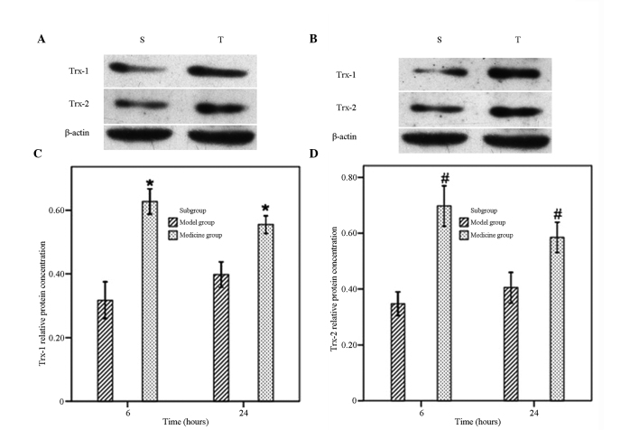
The aims of the present study were to investigate the protective effect of tanshinone IIA on the brain and its therapeutic time window in a rat model of cerebral ischemia-reperfusion. The rat model of cerebral ischemia-reperfusion was established by suture occlusion. In an initial experiment, male Sprague-Dawley (SD) rats were randomly divided into control cerebral ischemia-reperfusion rat model, tanshinone IIA1 (TSA1), tanshinone IIA4 (TSA4), tanshinone IIA6 (TSA6) and tanshinone IIA12 (TSA12) groups (n=8 per group). The rats in the control group were given 4 ml phosphate-buffered saline (PBS) intraperitoneally following suture occlusion. The other groups were respectively treated with 25 mg/kg tanshinone IIA intraperitoneally at 1, 4, 6 and 12 h following the initiation of reperfusion and once a day for a total of three days. The grades of neurologic impairment and volume of cerebral infarction of each group were measured 72 h after suture occlusion. In another experiment, 16 male SD rats were randomly divided into a 6 h reperfusion group and a 24 h reperfusion group following drug administration. The rats in each group were further divided into a control subgroup (4 ml PBS) and a tanshinone IIA subgroup (25 mg/kg). The rats were immediately administered their respective treatments following the establishment of the model. The rats were decapitated 6 and 24 h after the initiation of reperfusion. The expression levels of cytoplasmic thioredoxin (Trx-1) and mitochondrial thioredoxin (Trx-2) in the ischemic penumbra were determined by western blot analysis. The nitric oxide (NO) levels, and total NO synthase (tNOS) and inducible NO synthase (iNOS) activities in the rat blood were measured using a reagent kit. The changes in cerebral blood flow were evaluated by Doppler imaging. The grade of neurological impairment of the TSA1 group was statistically lower than that of the other groups (P<0.05). The cerebral infarction volume results showed that the volumes of infarction in the TSA1 and TSA4 groups were lower than those in the other groups (P<0.05). Tanshinone IIA significantly increased cerebral blood flow compared with that of the control group (P<0.05). Moreover, tanshinone IIA significantly increased the expression levels of Trx-1 and Trx-2 compared with those in the control group (P<0.05). Tanshinone IIA significantly decreased the NO levels and iNOS and tNOS activities compared with those of the control group (P<0.05). However, the iNOS activity in the rats in the 6 h reperfusion group was not statistically significantly different from that of the respective control group (P>0.05). Tanshinone IIA has a protective effect on the cranial nerves when administered during the initial stages of cerebral ischemia. This protective effect is associated with an improvement of cerebral blood flow as well as an increase in anti-oxygen radical and anti-inflammatory activities.
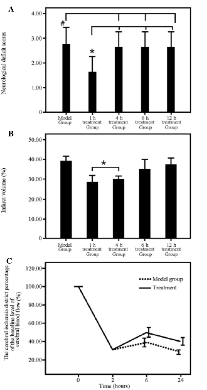
本研究旨在探讨丹参酮IIA对脑缺血再灌注大鼠模型的脑保护作用及其治疗时间窗。采用线栓法建立大鼠脑缺血再灌注模型。在初步实验中,将雄性Sprague-Dawley(SD)大鼠随机分为对照组(脑缺血再灌注大鼠模型)、丹参酮IIA 1小时给药组(TSA1)、丹参酮IIA 4小时给药组(TSA4)、丹参酮IIA 6小时给药组(TSA6)和丹参酮IIA 12小时给药组(TSA12)(每组n = 8)。对照组大鼠在结扎后腹腔注射4 ml磷酸盐缓冲盐水(PBS)。其他组在再灌注开始后1、4、6和12小时分别腹腔注射25 mg/kg丹参酮IIA,每天1次,共3天。结扎后72小时测量每组的神经功能缺损评分和脑梗死体积。在另一实验中,16只雄性SD大鼠在给药后随机分为6小时再灌注组和24小时再灌注组。每组大鼠进一步分为对照组(4 ml PBS)和丹参酮IIA组(25 mg/kg)。模型建立后立即给予相应处理。再灌注开始后6小时和24小时断头处死大鼠。采用蛋白质免疫印迹分析测定缺血半暗带中细胞质硫氧还蛋白(Trx-1)和线粒体硫氧还蛋白(Trx-2)的表达水平。使用试剂盒测量大鼠血液中的一氧化氮(NO)水平、总一氧化氮合酶(tNOS)和诱导型一氧化氮合酶(iNOS)活性。通过多普勒成像评估脑血流量变化。TSA1组的神经功能缺损评分在统计学上低于其他组(P<0.05)。脑梗死体积结果显示,TSA1组和TSA4组的梗死体积低于其他组(P<0.05)。与对照组相比,丹参酮IIA显著增加脑血流量(P<0.05)。此外,与对照组相比,丹参酮IIA显著增加Trx-1和Trx-2的表达水平(P<0.05)。与对照组相比,丹参酮IIA显著降低NO水平、iNOS和tNOS活性(P<0.05)。然而,6小时再灌注组大鼠的iNOS活性与相应对照组相比无统计学差异(P>0.05)。在脑缺血初期给予丹参酮IIA对脑神经具有保护作用。这种保护作用与脑血流量的改善以及抗氧化自由基和抗炎活性的增加有关。