Jalali Mir Mohammad, Gerami Hooshang, Rahimi Abbas, Jafari Manizheh
Department of Otorhinolaryngology, Amiralmomenin Hospital, Guilan University of Medical Sciences, Guilan, Iran.
Department of Radiotherapy, Razi Hospital, Guilan University of Medical Sciences, Guilan, Iran.
Iran J Otorhinolaryngol. 2014 Oct;26(77):211-7.
Radiotherapy is a common treatment modality for patients with head and neck malignancies. As the nose lies within the field of radiotherapy of the head and neck, the olfactory fibers and olfactory receptors may be affected by radiation. The aim of this study was to evaluate changes in olfactory threshold in patients with head and neck malignancies who have received radiation to the head and neck.
The olfactory threshold of patients with head and neck malignancies was assessed prospectively before radiation therapy and serially for up to 6 months after radiotherapy using sniff bottles. In vivo dosimetry was performed using 82 LiF (MCP) chips and a thermoluminescent dosimeter (TLD) system.
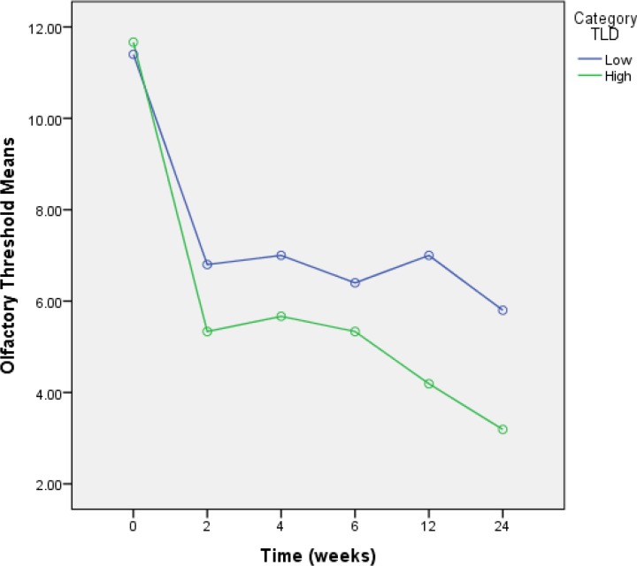
Sixty-one patients were recruited before radiotherapy was commenced. Seven patients did not return for evaluation after radiation. Fifty-four patients were available for follow-up assessment (28 women, 26 men; age, 22-86 years; median, 49 years). Total radiation dose was 50.1 Gy (range, 30-66 Gy). Mean olfactory threshold scores were found to deteriorate significantly at various timepoints after radiotherapy (11.7 before radiotherapy versus 4.0 at Month 6, general linear model, P<0.0001). With in vivo dosimetry, we found that the median measured dose to the olfactory area was 334 µC. We also identified a cutoff point according to the dose to the olfactory epithelium. Olfactory threshold was significantly decreased 2-6 weeks after initiation of therapy, with cumulative local radiation >135 µC (Mann-Whitney U test, P=0.01).
Deterioration in olfactory threshold scores was found at 6 months after initiation of radiation therapy. Provided that these results are reproducible, an evaluation of olfactory functioning in patients with head and neck malignancies using in vivo dosimetry may be useful for determining the optimal dose for patients treated with conformal radiotherapy techniques while avoiding the side effects of radiation.
放射治疗是头颈部恶性肿瘤患者的常见治疗方式。由于鼻子位于头颈部放射治疗区域内,嗅觉纤维和嗅觉受体可能会受到辐射影响。本研究的目的是评估接受过头颈部放疗的头颈部恶性肿瘤患者嗅觉阈值的变化。
对头颈部恶性肿瘤患者在放疗前进行前瞻性嗅觉阈值评估,并在放疗后连续6个月使用嗅瓶进行评估。使用82LiF(MCP)芯片和热释光剂量计(TLD)系统进行体内剂量测定。
放疗开始前招募了61名患者。7名患者放疗后未返回进行评估。54名患者可供随访评估(28名女性,26名男性;年龄22 - 86岁;中位数49岁)。总辐射剂量为50.1 Gy(范围30 - 66 Gy)。发现放疗后不同时间点的平均嗅觉阈值评分显著恶化(放疗前为11.7,第6个月时为4.0,一般线性模型,P<0.0001)。通过体内剂量测定,我们发现嗅觉区域的中位测量剂量为334 µC。我们还根据嗅觉上皮的剂量确定了一个临界点。治疗开始后2 - 6周,当累积局部辐射>135 µC时,嗅觉阈值显著降低(曼 - 惠特尼U检验,P = 0.01)。
放疗开始后6个月发现嗅觉阈值评分恶化。如果这些结果具有可重复性,那么在头颈部恶性肿瘤患者中使用体内剂量测定评估嗅觉功能,可能有助于确定采用适形放疗技术治疗患者的最佳剂量,同时避免辐射副作用。