Czapla Norbert, Lokaj Marek, Falkowski Aleksander, Prowans Piotr
Clinic of Plastic, Endocrine and General Surgery, Pomeranian Medical University, Police, Poland.
Department of Interventional Radiology, Pomeranian Medical University, Szczecin, Poland.
Wideochir Inne Tech Maloinwazyjne. 2014 Sep;9(3):319-28. doi: 10.5114/wiitm.2014.44056. Epub 2014 Jul 10.
Methods allowing one to locate the position of a cutaneous perforator do not allow one to determine the boundaries of the vascularized skin. In clinical practice this causes complications in the form of marginal necrosis of the flap.
To examine the usefulness of thermography to assess the extent of vascularization of the skin and subcutaneous tissue by a single perforator.
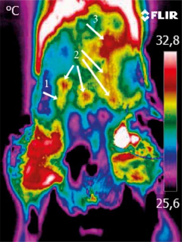
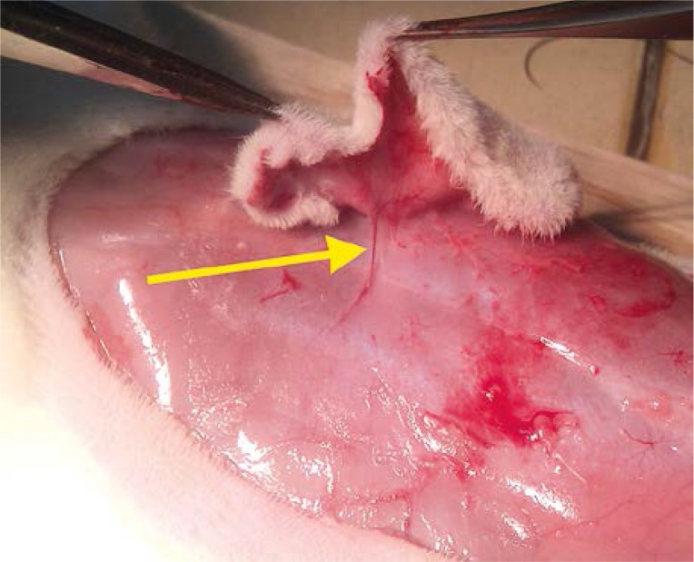
Thirty-one male rats were used. Using dynamic thermography the perforators on the abdominal skin were located. Afterwards the flap was prepared on a randomly chosen perforator. After 24 h the extent of vascularization of the skin by a single perforator was examined.
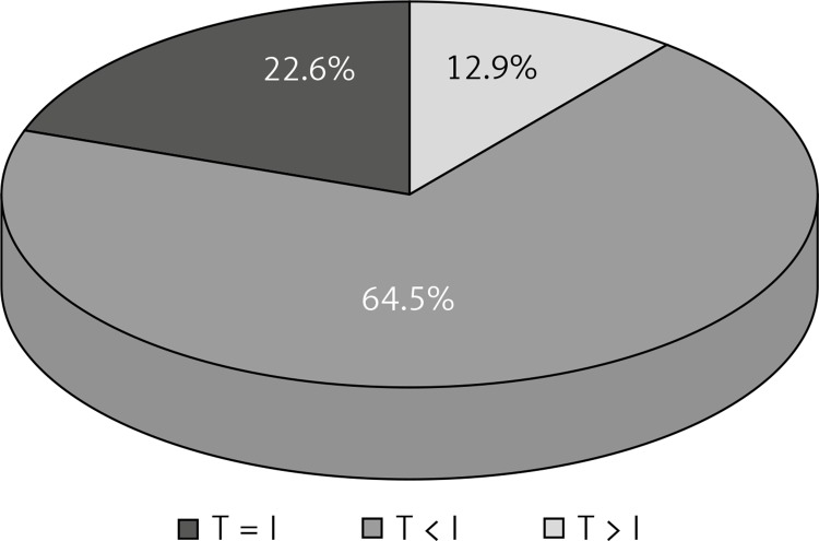
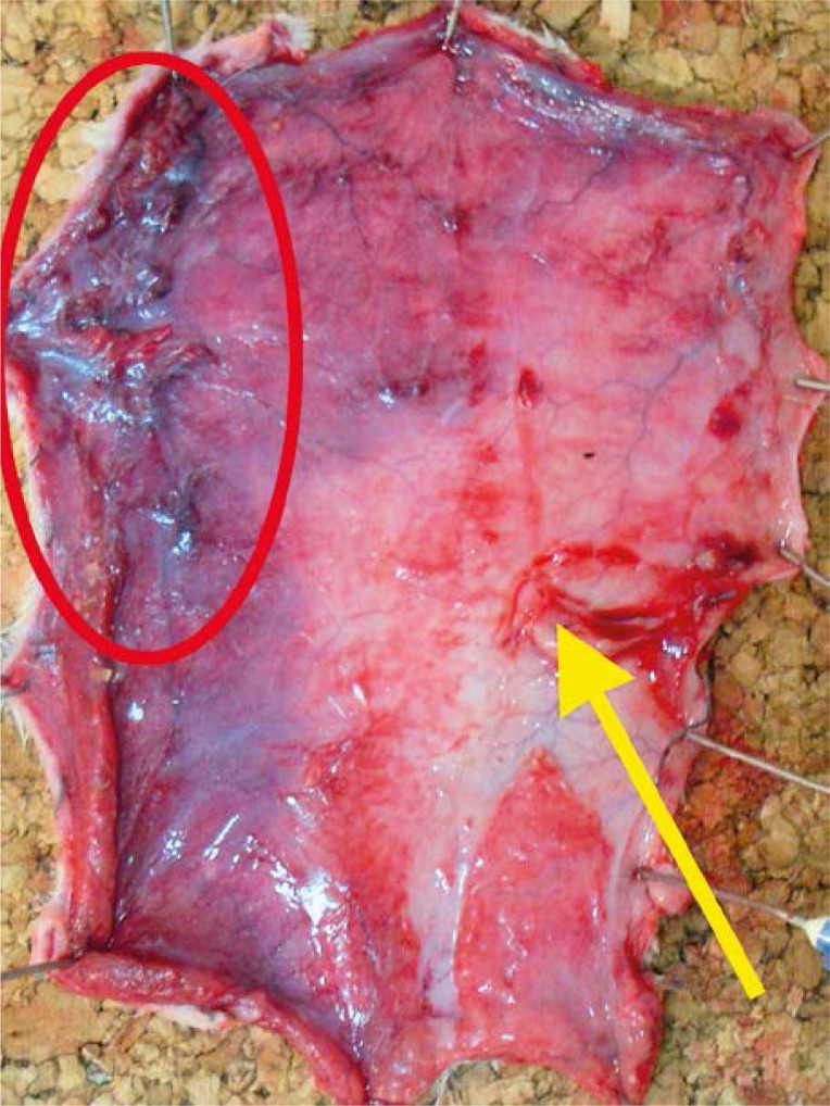
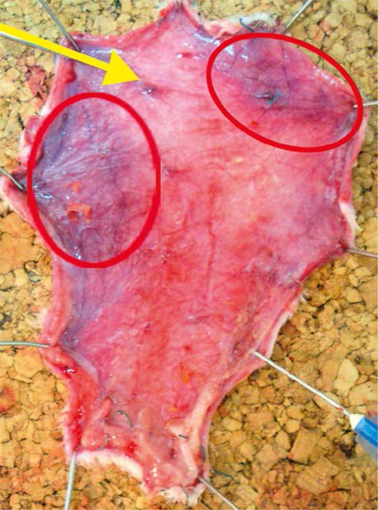
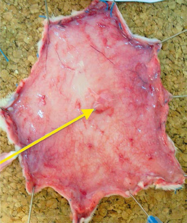
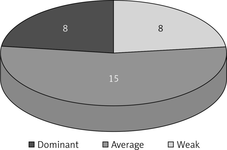
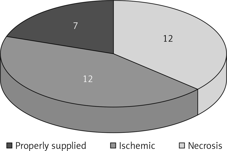
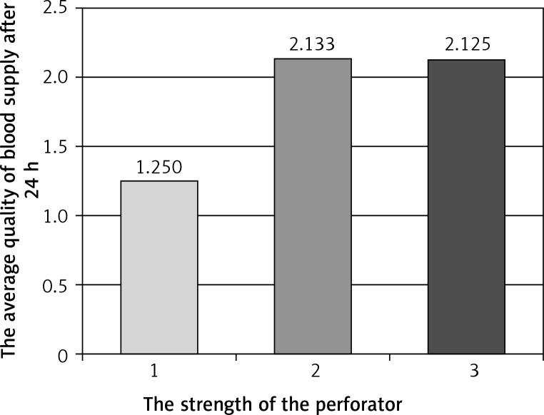
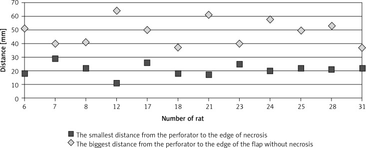
In 22.5% of cases the number of perforators marked in the thermography was equal to the number of perforators marked intraoperatively, in 64.5% it was lower and in 13% higher. The use of thermography has shown that basing the flap vascularization on the perforator with low efficiency resulted in statistically more frequent occurrence of ischemia and partial necrosis of the flap (p = 0.024). Partial necrosis of the flap occurred in 12 of 31 cases, always in the area in which during the preoperative thermography no perforators were found. The areas of necrosis occurred irrespectively of the distance from the supplying vessel.
When designing the shape of the flap, the distribution of all perforators must be considered. The perforators need to be included in the area of prepared tissues because their location indicates the area with a more efficient network of vessels.
现有的确定皮肤穿支位置的方法无法确定血管化皮肤的边界。在临床实践中,这会导致皮瓣边缘坏死等并发症。
研究热成像技术在评估单个穿支对皮肤和皮下组织血管化程度方面的实用性。
使用31只雄性大鼠。通过动态热成像定位腹部皮肤的穿支。之后在随机选择的一个穿支上制备皮瓣。24小时后,检查单个穿支对皮肤的血管化程度。
在22.5%的病例中,热成像标记的穿支数量与术中标记的穿支数量相等,64.5%的病例中热成像标记的穿支数量较少,13%的病例中热成像标记的穿支数量较多。热成像技术的应用表明,基于低效穿支进行皮瓣血管化在统计学上导致皮瓣缺血和部分坏死的发生率更高(p = 0.024)。31例中有12例发生皮瓣部分坏死,坏死部位均为术前热成像未发现穿支的区域。坏死区域与供血血管的距离无关。
在设计皮瓣形状时,必须考虑所有穿支的分布。穿支应包含在准备组织的区域内,因为它们的位置表明该区域血管网络更有效。