• 文献检索
  • 文档翻译
  • 深度研究
  • 学术资讯
  • Suppr Zotero 插件Zotero 插件
  • 邀请有礼
  • 套餐&价格
  • 历史记录
应用&插件
Suppr Zotero 插件Zotero 插件浏览器插件Mac 客户端Windows 客户端微信小程序
定价
高级版会员购买积分包购买API积分包
服务
文献检索文档翻译深度研究API 文档MCP 服务
关于我们
关于 Suppr公司介绍联系我们用户协议隐私条款
关注我们

Suppr 超能文献

核心技术专利:CN118964589B侵权必究
粤ICP备2023148730 号-1Suppr @ 2026

文献检索

告别复杂PubMed语法,用中文像聊天一样搜索,搜遍4000万医学文献。AI智能推荐,让科研检索更轻松。

立即免费搜索

文件翻译

保留排版,准确专业,支持PDF/Word/PPT等文件格式,支持 12+语言互译。

免费翻译文档

深度研究

AI帮你快速写综述,25分钟生成高质量综述,智能提取关键信息,辅助科研写作。

立即免费体验

内胚层在肝脏发育过程中产生内皮细胞。

Endoderm generates endothelial cells during liver development.

机构信息

Department of Developmental and Regenerative Biology, Black Family Stem Cell Institute, Icahn School of Medicine, Mount Sinai, New York, NY 10029, USA.

INSERM U972, Hospital Paul Brousse, 12 Avenue Paul Vaillant Couturier, 94807 Villejuif, France.

出版信息

Stem Cell Reports. 2014 Oct 14;3(4):556-65. doi: 10.1016/j.stemcr.2014.08.009. Epub 2014 Sep 18.

DOI:10.1016/j.stemcr.2014.08.009
PMID:25358784
原文链接:https://pmc.ncbi.nlm.nih.gov/articles/PMC4223703/
Abstract

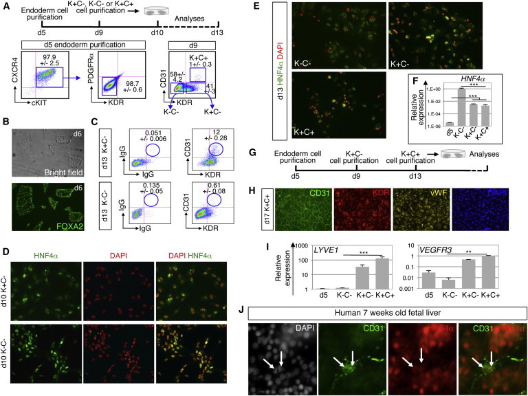
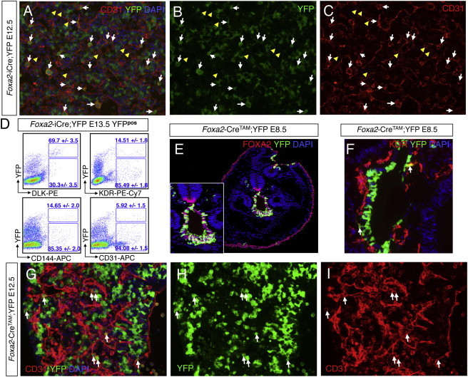
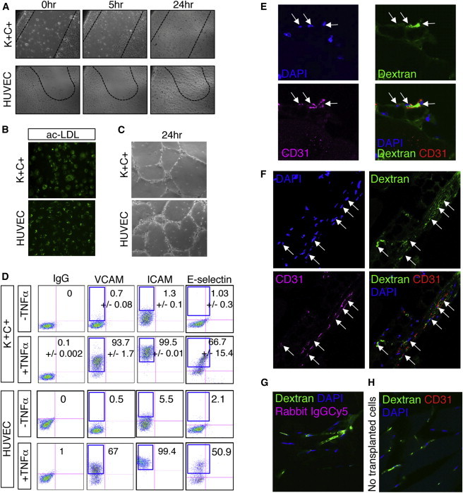
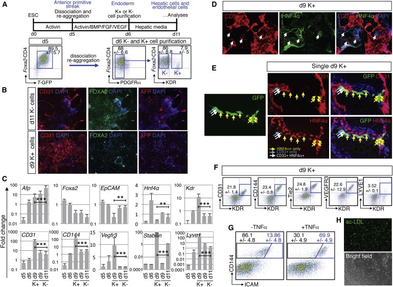
Organogenesis requires expansion of the embryonic vascular plexus that migrates into developing organs through a process called angiogenesis. Mesodermal progenitors are thought to derive endothelial cells (ECs) that contribute to both embryonic vasculogenesis and the subsequent organ angiogenesis. Here, we demonstrate that during development of the liver, which is an endoderm derivative, a subset of ECs is generated from FOXA2+ endoderm-derived fetal hepatoblast progenitor cells expressing KDR (VEGFR2/FLK-1). Using human and mouse embryonic stem cell models, we demonstrate that KDR+FOXA2+ endoderm cells developing in hepatic differentiation cultures generate functional ECs. This introduces the concept that ECs originate not exclusively from mesoderm but also from endoderm, supported in Foxa2 lineage-tracing mouse embryos by the identification of FOXA2+ cell-derived CD31+ ECs that integrate the vascular network of developing fetal livers.

摘要

器官发生需要胚胎血管丛的扩张,该血管丛通过称为血管生成的过程迁移到正在发育的器官中。中胚层祖细胞被认为来源于内皮细胞(ECs),这些细胞有助于胚胎血管发生和随后的器官血管生成。在这里,我们证明在肝脏的发育过程中,肝脏是内胚层的衍生物,一部分 ECs 是由表达 KDR(VEGFR2/FLK-1)的 FOXA2+内胚层来源的胎儿肝祖细胞产生的。使用人和小鼠胚胎干细胞模型,我们证明在肝分化培养中发育的 KDR+FOXA2+内胚层细胞可产生功能性 ECs。这引入了一个概念,即 ECs 不仅源自中胚层,而且还源自内胚层,在 Foxa2 谱系追踪小鼠胚胎中得到支持,因为鉴定出 FOXA2+细胞衍生的 CD31+ECs 整合了发育中胎儿肝脏的血管网络。

https://cdn.ncbi.nlm.nih.gov/pmc/blobs/5200/4223703/87b9d23b39f2/gr4.jpg
https://cdn.ncbi.nlm.nih.gov/pmc/blobs/5200/4223703/d3126f6bf84b/gr1.jpg
https://cdn.ncbi.nlm.nih.gov/pmc/blobs/5200/4223703/83bd0d995846/gr2.jpg
https://cdn.ncbi.nlm.nih.gov/pmc/blobs/5200/4223703/589959b9002f/gr3.jpg
https://cdn.ncbi.nlm.nih.gov/pmc/blobs/5200/4223703/87b9d23b39f2/gr4.jpg
https://cdn.ncbi.nlm.nih.gov/pmc/blobs/5200/4223703/d3126f6bf84b/gr1.jpg
https://cdn.ncbi.nlm.nih.gov/pmc/blobs/5200/4223703/83bd0d995846/gr2.jpg
https://cdn.ncbi.nlm.nih.gov/pmc/blobs/5200/4223703/589959b9002f/gr3.jpg
https://cdn.ncbi.nlm.nih.gov/pmc/blobs/5200/4223703/87b9d23b39f2/gr4.jpg

相似文献

1
Endoderm generates endothelial cells during liver development.内胚层在肝脏发育过程中产生内皮细胞。
Stem Cell Reports. 2014 Oct 14;3(4):556-65. doi: 10.1016/j.stemcr.2014.08.009. Epub 2014 Sep 18.
2
An endothelial cell niche induces hepatic specification through dual repression of Wnt and Notch signaling.内皮细胞龛通过双重抑制 Wnt 和 Notch 信号诱导肝特化。
Stem Cells. 2011 Feb;29(2):217-28. doi: 10.1002/stem.576.
3
miR-335 promotes mesendodermal lineage segregation and shapes a transcription factor gradient in the endoderm.miR-335 促进中胚层和内胚层的谱系分离,并在内胚层中形成转录因子梯度。
Development. 2014 Feb;141(3):514-25. doi: 10.1242/dev.104232.
4
ROCK suppression promotes differentiation and expansion of endothelial cells from embryonic stem cell-derived Flk1(+) mesodermal precursor cells.ROCK 抑制促进了胚胎干细胞来源的 Flk1(+)中胚层前体细胞向血管内皮细胞的分化和扩增。
Blood. 2012 Sep 27;120(13):2733-44. doi: 10.1182/blood-2012-04-421610. Epub 2012 Aug 14.
5
Enforced expression of Mixl1 during mouse ES cell differentiation suppresses hematopoietic mesoderm and promotes endoderm formation.在小鼠胚胎干细胞分化过程中强制表达Mixl1可抑制造血中胚层并促进内胚层形成。
Stem Cells. 2009 Feb;27(2):363-74. doi: 10.1634/stemcells.2008-1008.
6
Endothelial cells instruct liver specification of embryonic stem cell-derived endoderm through endothelial VEGFR2 signaling and endoderm epigenetic modifications.内皮细胞通过内皮血管内皮生长因子受体2(VEGFR2)信号传导和内胚层表观遗传修饰指导胚胎干细胞来源的内胚层向肝脏方向分化。
Stem Cell Res. 2018 Jul;30:163-170. doi: 10.1016/j.scr.2018.06.004. Epub 2018 Jun 21.
7
Soluble Flt-1 regulates Flk-1 activation to control hematopoietic and endothelial development in an oxygen-responsive manner.可溶性Flt-1以氧反应性方式调节Flk-1激活,以控制造血和内皮发育。
Stem Cells. 2008 Nov;26(11):2832-42. doi: 10.1634/stemcells.2008-0237. Epub 2008 Sep 4.
8
Multiple mesoderm subsets give rise to endothelial cells, whereas hematopoietic cells are differentiated only from a restricted subset in embryonic stem cell differentiation culture.多个中胚层亚群可产生内皮细胞,而造血细胞仅在胚胎干细胞分化培养中从一个受限亚群分化而来。
Stem Cells. 2008 Feb;26(2):401-11. doi: 10.1634/stemcells.2006-0809. Epub 2007 Nov 8.
9
KDR identifies a conserved human and murine hepatic progenitor and instructs early liver development.KDR 鉴定出一个保守的人类和鼠类肝祖细胞,并指导早期肝脏发育。
Cell Stem Cell. 2013 Jun 6;12(6):748-60. doi: 10.1016/j.stem.2013.04.026.
10
Profiling of transcriptional and epigenetic changes during directed endothelial differentiation of human embryonic stem cells identifies FOXA2 as a marker of early mesoderm commitment.对人类胚胎干细胞定向内皮分化过程中转录和表观遗传变化的分析确定FOXA2为早期中胚层定向的标志物。
Stem Cell Res Ther. 2013 Apr 24;4(2):36. doi: 10.1186/scrt192.

引用本文的文献

1
Single-cell spatial mapping reveals reproducible cell type organization and spatially-dependent gene expression in gastruloids.单细胞空间图谱揭示了原肠胚样结构中可重复的细胞类型组织和空间依赖性基因表达。
bioRxiv. 2025 Jul 31:2025.07.14.664617. doi: 10.1101/2025.07.14.664617.
2
hESCs-derived Organoids Achieve Liver Zonation Features through LSEC Modulation.人胚胎干细胞来源的类器官通过肝窦内皮细胞调节实现肝脏分区特征。
Adv Sci (Weinh). 2025 May;12(20):e2411667. doi: 10.1002/advs.202411667. Epub 2025 Apr 25.
3
Exploring vasculogenesis in the normal human kidney and clear cell renal cell carcinoma: insights from development to tumor progression and biomarkers for therapy response.

本文引用的文献

1
Molecular signatures of tissue-specific microvascular endothelial cell heterogeneity in organ maintenance and regeneration.组织特异性微血管内皮细胞异质性在器官维持和再生中的分子特征。
Dev Cell. 2013 Jul 29;26(2):204-19. doi: 10.1016/j.devcel.2013.06.017. Epub 2013 Jul 18.
2
KDR identifies a conserved human and murine hepatic progenitor and instructs early liver development.KDR 鉴定出一个保守的人类和鼠类肝祖细胞,并指导早期肝脏发育。
Cell Stem Cell. 2013 Jun 6;12(6):748-60. doi: 10.1016/j.stem.2013.04.026.
3
Mind bomb 1 is required for pancreatic β-cell formation.
探索正常人类肾脏和透明细胞肾细胞癌中的血管生成:从发育到肿瘤进展的见解以及治疗反应的生物标志物
Front Oncol. 2024 Apr 30;14:1375190. doi: 10.3389/fonc.2024.1375190. eCollection 2024.
4
Liver specification of human iPSC-derived endothelial cells transplanted into mouse liver.移植到小鼠肝脏中的人诱导多能干细胞衍生内皮细胞向肝脏的定向分化
JHEP Rep. 2024 Feb 1;6(5):101023. doi: 10.1016/j.jhepr.2024.101023. eCollection 2024 May.
5
Single-nucleus transcriptome sequencing reveals hepatic cell atlas in pigs.单细胞转录组测序揭示猪的肝实质细胞图谱。
BMC Genomics. 2023 Dec 12;24(1):770. doi: 10.1186/s12864-023-09765-9.
6
Clonal and Scalable Endothelial Progenitor Cell Lines from Human Pluripotent Stem Cells.源自人多能干细胞的克隆且可扩展的内皮祖细胞系。
Biomedicines. 2023 Oct 13;11(10):2777. doi: 10.3390/biomedicines11102777.
7
Organ-Specific Endothelial Cell Differentiation and Impact of Microenvironmental Cues on Endothelial Heterogeneity.器官特异性内皮细胞分化和微环境线索对内皮细胞异质性的影响。
Int J Mol Sci. 2022 Jan 27;23(3):1477. doi: 10.3390/ijms23031477.
8
Engineering the Vasculature of Stem-Cell-Derived Liver Organoids.工程化干细胞衍生肝类器官的脉管系统。
Biomolecules. 2021 Jun 30;11(7):966. doi: 10.3390/biom11070966.
9
Analyses of Avascular Mutants Reveal Unique Transcriptomic Signature of Non-conventional Endothelial Cells.无血管突变体分析揭示非常规内皮细胞独特的转录组特征。
Front Cell Dev Biol. 2020 Nov 23;8:589717. doi: 10.3389/fcell.2020.589717. eCollection 2020.
10
A human cell atlas of fetal chromatin accessibility.人类胚胎染色质可及性细胞图谱。
Science. 2020 Nov 13;370(6518). doi: 10.1126/science.aba7612.
脑炸弹 1 对于胰腺 β 细胞的形成是必需的。
Proc Natl Acad Sci U S A. 2012 May 8;109(19):7356-61. doi: 10.1073/pnas.1203605109. Epub 2012 Apr 23.
4
An endothelial cell niche induces hepatic specification through dual repression of Wnt and Notch signaling.内皮细胞龛通过双重抑制 Wnt 和 Notch 信号诱导肝特化。
Stem Cells. 2011 Feb;29(2):217-28. doi: 10.1002/stem.576.
5
Inductive angiocrine signals from sinusoidal endothelium are required for liver regeneration.窦状内皮细胞的诱导性血管分泌信号对于肝再生是必需的。
Nature. 2010 Nov 11;468(7321):310-5. doi: 10.1038/nature09493.
6
Sinusoid development and morphogenesis may be stimulated by VEGF-Flk-1 signaling during fetal mouse liver development.在胎鼠肝发育过程中,VEGF-Flk-1 信号可能刺激窦状隙发育和形态发生。
Dev Dyn. 2010 Feb;239(2):386-97. doi: 10.1002/dvdy.22162.
7
A boost of BMP4 accelerates the commitment of human embryonic stem cells to the endothelial lineage.BMP4 的促进作用加速了人胚胎干细胞向血管内皮谱系的定向分化。
Stem Cells. 2009 Aug;27(8):1750-9. doi: 10.1002/stem.100.
8
Embryology of extra- and intrahepatic bile ducts, the ductal plate.肝外和肝内胆管的胚胎学,导管板
Anat Rec (Hoboken). 2008 Jun;291(6):628-35. doi: 10.1002/ar.20710.
9
Human cardiovascular progenitor cells develop from a KDR+ embryonic-stem-cell-derived population.人类心血管祖细胞源自KDR⁺胚胎干细胞衍生群体。
Nature. 2008 May 22;453(7194):524-8. doi: 10.1038/nature06894. Epub 2008 Apr 23.
10
System for tamoxifen-inducible expression of cre-recombinase from the Foxa2 locus in mice.用于在小鼠中从Foxa2基因座进行他莫昔芬诱导型Cre重组酶表达的系统。
Dev Dyn. 2008 Feb;237(2):447-53. doi: 10.1002/dvdy.21415.