Suarez Ralph O, Taimouri Vahid, Boyer Katrina, Vega Clemente, Rotenberg Alexander, Madsen Joseph R, Loddenkemper Tobias, Duffy Frank H, Prabhu Sanjay P, Warfield Simon K
Department of Radiology, Boston Children's Hospital, Harvard Medical School, Boston, MA, USA.
Department of Radiology, Boston Children's Hospital, Harvard Medical School, Boston, MA, USA.
Epilepsy Res. 2014 Dec;108(10):1874-88. doi: 10.1016/j.eplepsyres.2014.09.016. Epub 2014 Sep 28.
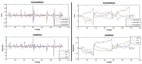
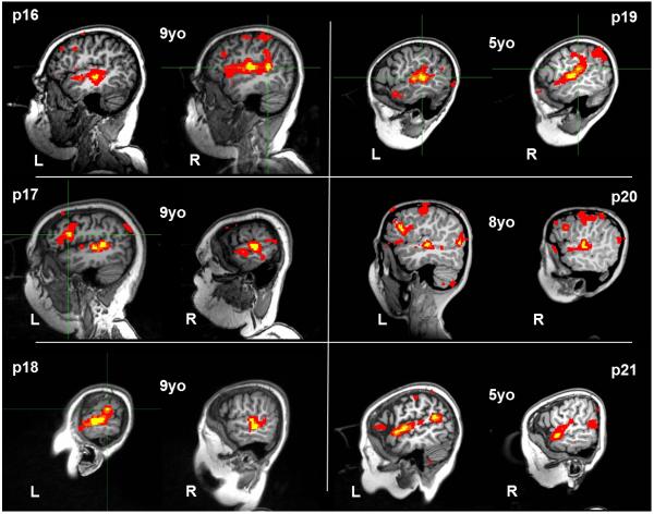
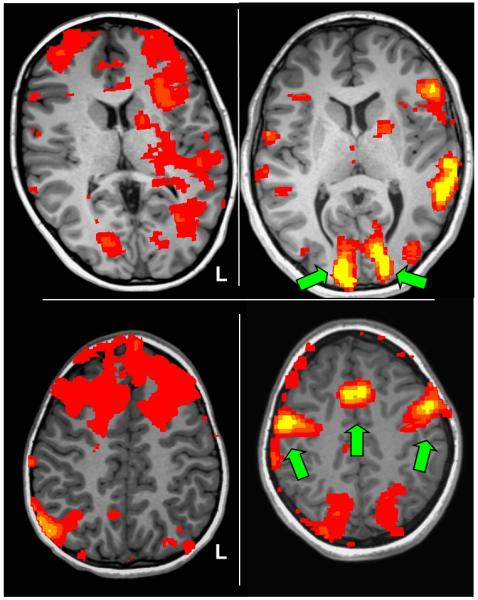
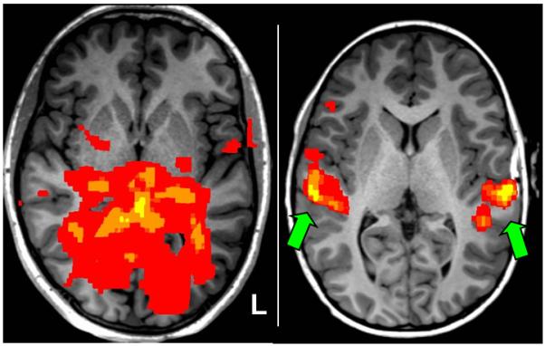
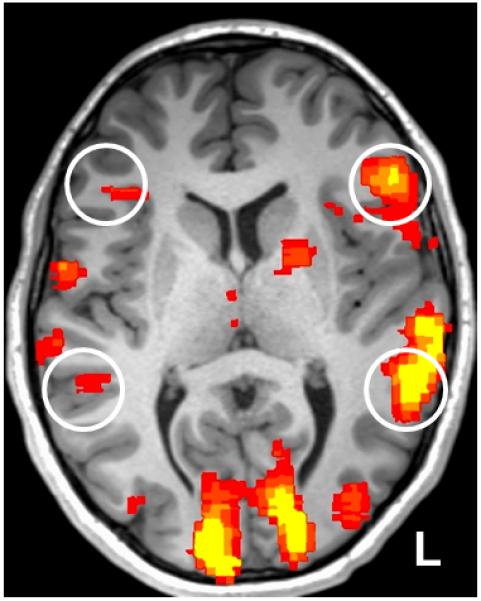
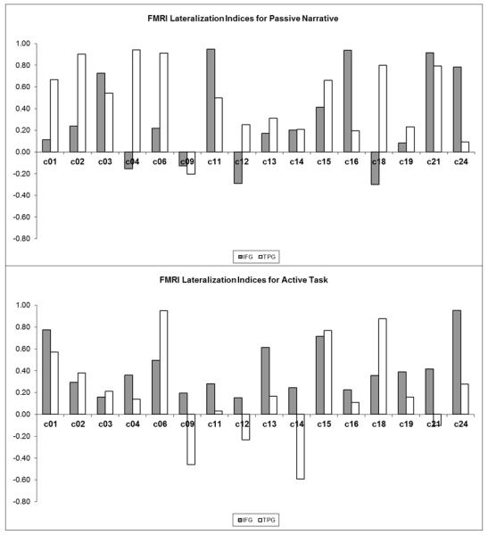
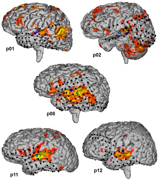
In this study we validate passive language fMRI protocols designed for clinical application in pediatric epilepsy surgical planning as they do not require overt participation from patients. We introduced a set of quality checks that assess reliability of noninvasive fMRI mappings utilized for clinical purposes. We initially compared two fMRI language mapping paradigms, one active in nature (requiring participation from the patient) and the other passive in nature (requiring no participation from the patient). Group-level analysis in a healthy control cohort demonstrated similar activation of the putative language centers of the brain in the inferior frontal (IFG) and temporoparietal (TPG) regions. Additionally, we showed that passive language fMRI produced more left-lateralized activation in TPG (LI=+0.45) compared to the active task; with similarly robust left-lateralized IFG (LI=+0.24) activations using the passive task. We validated our recommended fMRI mapping protocols in a cohort of 15 pediatric epilepsy patients by direct comparison against the invasive clinical gold-standards. We found that language-specific TPG activation by fMRI agreed to within 9.2mm to subdural localizations by invasive functional mapping in the same patients, and language dominance by fMRI agreed with Wada test results at 80% congruency in TPG and 73% congruency in IFG. Lastly, we tested the recommended passive language fMRI protocols in a cohort of very young patients and confirmed reliable language-specific activation patterns in that challenging cohort. We concluded that language activation maps can be reliably achieved using the passive language fMRI protocols we proposed even in very young (average 7.5 years old) or sedated pediatric epilepsy patients.
在本研究中,我们验证了专为小儿癫痫手术规划临床应用设计的被动语言功能磁共振成像(fMRI)方案,因为这些方案无需患者的主动参与。我们引入了一组质量检查,以评估用于临床目的的非侵入性fMRI映射的可靠性。我们最初比较了两种fMRI语言映射范式,一种是主动型(需要患者参与),另一种是被动型(无需患者参与)。对健康对照队列的组水平分析表明,大脑假定语言中枢在额下回(IFG)和颞顶叶(TPG)区域有相似的激活。此外,我们发现与主动任务相比,被动语言fMRI在TPG区域产生了更多的左侧化激活(LI = +0.45);使用被动任务时,IFG也有同样强烈的左侧化激活(LI = +0.24)。我们通过与侵入性临床金标准直接比较,在15名小儿癫痫患者队列中验证了我们推荐的fMRI映射方案。我们发现,fMRI显示的特定语言TPG激活与同一患者侵入性功能映射的硬膜下定位在9.2毫米范围内一致,fMRI显示的语言优势与Wada测试结果在TPG区域的一致性为80%,在IFG区域为73%。最后,我们在一组非常年幼的患者中测试了推荐的被动语言fMRI方案,并在这个具有挑战性的队列中证实了可靠的特定语言激活模式。我们得出结论,即使在非常年幼(平均7.5岁)或接受镇静的小儿癫痫患者中,使用我们提出的被动语言fMRI方案也能可靠地获得语言激活图。