• 文献检索
  • 文档翻译
  • 深度研究
  • 学术资讯
  • Suppr Zotero 插件Zotero 插件
  • 邀请有礼
  • 套餐&价格
  • 历史记录
应用&插件
Suppr Zotero 插件Zotero 插件浏览器插件Mac 客户端Windows 客户端微信小程序
定价
高级版会员购买积分包购买API积分包
服务
文献检索文档翻译深度研究API 文档MCP 服务
关于我们
关于 Suppr公司介绍联系我们用户协议隐私条款
关注我们

Suppr 超能文献

核心技术专利:CN118964589B侵权必究
粤ICP备2023148730 号-1Suppr @ 2026

文献检索

告别复杂PubMed语法,用中文像聊天一样搜索,搜遍4000万医学文献。AI智能推荐,让科研检索更轻松。

立即免费搜索

文件翻译

保留排版,准确专业,支持PDF/Word/PPT等文件格式,支持 12+语言互译。

免费翻译文档

深度研究

AI帮你快速写综述,25分钟生成高质量综述,智能提取关键信息,辅助科研写作。

立即免费体验

使用眼前节光学相干断层扫描和共聚焦显微镜对两种不同方案的加速角膜胶原交联术后角膜基质分界线的评估。

Evaluation of corneal stromal demarcation line after two different protocols of accelerated corneal collagen cross-linking procedures using anterior segment optical coherence tomography and confocal microscopy.

作者信息

Ozgurhan Engin Bilge, Sezgin Akcay Betul Ilkay, Yildirim Yusuf, Karatas Gonul, Kurt Tugba, Demirok Ahmet

机构信息

Department of Ophthalmology, Beyoglu Eye Training and Research Hospital, Bereketzade Camii Sok, Kuledibi, Beyoglu, 34421 Istanbul, Turkey.

Department of Ophthalmology, Umraniye Training and Research Hospital, Istanbul, Turkey.

出版信息

J Ophthalmol. 2014;2014:981893. doi: 10.1155/2014/981893. Epub 2014 Nov 18.

DOI:10.1155/2014/981893
PMID:25485145
原文链接:https://pmc.ncbi.nlm.nih.gov/articles/PMC4251080/
Abstract

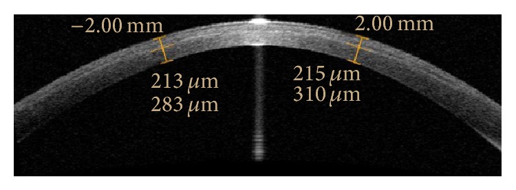
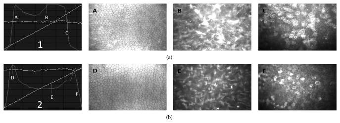
Purpose. To evaluate the depth of corneal stromal demarcation line using AS-OCT and confocal microscopy after two different protocols of accelerated corneal collagen cross-linking procedures (CXL). Methods. Patients with keratoconus were divided into two groups. Peschke CXL device (Peschke CCL-VARIO Meditrade GmbH) applied UVA light with an intended irradiance of 18.0 mW/cm(2) for 5 minutes after applying riboflavin for 20 minutes (group 1) and 30 minutes (group 2). One month postoperatively, corneal stromal demarcation line was measured using AS-OCT and confocal microscopy. Results. This study enrolled 34 eyes of 34 patients (17 eyes in group 1 and 17 eyes in group 2). The mean depth of the corneal stromal demarcation line was 208.64 ± 18.41 μm in group 1 and 240.37 ± 18.89 μm in group 2 measured with AS OCT, while it was 210.29 ± 18.66 μm in group 1 and 239.37 ± 20.07 μm in group 2 measured with confocal microscopy. Corneal stromal demarcation line depth measured with AS OCT or confocal microscopy was significantly deeper in group 2 than group 1 (P < 0.01). Conclusion. The group in which riboflavin was applied for 30 minutes showed significantly deeper corneal stromal demarcation line than the group in which riboflavin was applied for 20 minutes.

摘要

目的。在两种不同的加速角膜胶原交联程序(CXL)方案后,使用眼前节光学相干断层扫描(AS - OCT)和共聚焦显微镜评估角膜基质分界线的深度。方法。圆锥角膜患者被分为两组。在应用核黄素20分钟(第1组)和30分钟(第2组)后,使用Peschke CXL设备(Peschke CCL - VARIO Meditrade GmbH)以预期辐照度18.0 mW/cm²施加紫外线A光5分钟。术后1个月,使用AS - OCT和共聚焦显微镜测量角膜基质分界线。结果。本研究纳入了34例患者的34只眼(第1组17只眼,第2组17只眼)。用AS - OCT测量时,第1组角膜基质分界线的平均深度为208.64±18.41μm,第2组为240.37±18.89μm;用共聚焦显微镜测量时,第1组为210.29±18.66μm,第2组为239.37±20.07μm。用AS - OCT或共聚焦显微镜测量的角膜基质分界线深度在第2组显著深于第1组(P < 0.01)。结论。应用核黄素30分钟的组比应用核黄素20分钟的组角膜基质分界线明显更深。

https://cdn.ncbi.nlm.nih.gov/pmc/blobs/73ed/4251080/338057e1fd19/JOPH2014-981893.002.jpg
https://cdn.ncbi.nlm.nih.gov/pmc/blobs/73ed/4251080/74f2bea3e292/JOPH2014-981893.001.jpg
https://cdn.ncbi.nlm.nih.gov/pmc/blobs/73ed/4251080/338057e1fd19/JOPH2014-981893.002.jpg
https://cdn.ncbi.nlm.nih.gov/pmc/blobs/73ed/4251080/74f2bea3e292/JOPH2014-981893.001.jpg
https://cdn.ncbi.nlm.nih.gov/pmc/blobs/73ed/4251080/338057e1fd19/JOPH2014-981893.002.jpg

相似文献

1
Evaluation of corneal stromal demarcation line after two different protocols of accelerated corneal collagen cross-linking procedures using anterior segment optical coherence tomography and confocal microscopy.使用眼前节光学相干断层扫描和共聚焦显微镜对两种不同方案的加速角膜胶原交联术后角膜基质分界线的评估。
J Ophthalmol. 2014;2014:981893. doi: 10.1155/2014/981893. Epub 2014 Nov 18.
2
Corneal Stromal Demarcation Line Depth Following Standard and a Modified High Intensity Corneal Cross-linking Protocol.标准及改良高强度角膜交联方案后的角膜基质分界线深度
J Refract Surg. 2016 Apr;32(4):218-22. doi: 10.3928/1081597X-20160216-01.
3
Corneal stromal demarcation line after accelerated crosslinking using continuous and pulsed light.使用连续光和脉冲光加速交联后的角膜基质分界线
J Cataract Refract Surg. 2015 Nov;41(11):2546-51. doi: 10.1016/j.jcrs.2015.04.033.
4
Evaluation of corneal stromal demarcation line depth following standard and a modified-accelerated collagen cross-linking protocol.标准及改良加速胶原交联方案后角膜基质分界线深度的评估
Am J Ophthalmol. 2014 Oct;158(4):671-675.e1. doi: 10.1016/j.ajo.2014.07.005. Epub 2014 Jul 15.
5
Corneal stromal demarcation line after 4 protocols of corneal crosslinking in keratoconus determined with anterior segment optical coherence tomography.四种角膜交联方案治疗圆锥角膜后角膜基质分界线的眼前节光学相干断层扫描评估。
J Cataract Refract Surg. 2018 May;44(5):596-602. doi: 10.1016/j.jcrs.2018.02.017.
6
Correlation of the corneal collagen cross-linking demarcation line using confocal microscopy and anterior segment optical coherence tomography in keratoconic patients.应用共焦显微镜和眼前节光学相干断层扫描评估圆锥角膜患者角膜胶原交联分界线。
Am J Ophthalmol. 2014 Jan;157(1):110-115.e1. doi: 10.1016/j.ajo.2013.09.010. Epub 2013 Nov 5.
7
Visibility and Depth of the Stromal Demarcation Line After Corneal Collagen Cross-Linking Using Anterior Segment Optical Coherence Tomography: Comparison Between Isoosmolar and Hypoosmolar Riboflavin.使用眼前节光学相干断层扫描技术观察角膜胶原交联术后基质分界线的可视性和深度:等渗与低渗核黄素的比较
Cornea. 2018 May;37(5):567-573. doi: 10.1097/ICO.0000000000001493.
8
Corneal stroma demarcation line after standard and high-intensity collagen crosslinking determined with anterior segment optical coherence tomography.使用眼前节光学相干断层扫描确定标准和高强度胶原交联后的角膜基质分界线。
J Cataract Refract Surg. 2014 May;40(5):736-40. doi: 10.1016/j.jcrs.2013.10.029. Epub 2014 Mar 12.
9
Comparison of the Central and Peripheral Corneal Stromal Demarcation Line Depth in Conventional Versus Accelerated Collagen Cross-Linking.传统与加速胶原交联中中央与周边角膜基质分界线深度的比较
Cornea. 2015 Nov;34(11):1432-6. doi: 10.1097/ICO.0000000000000626.
10
Evaluation of the corneal collagen cross-linking demarcation line profile using anterior segment optical coherence tomography.使用眼前节光学相干断层扫描评估角膜胶原交联分界线轮廓。
Cornea. 2013 Jul;32(7):907-10. doi: 10.1097/ICO.0b013e31828733ea.

引用本文的文献

1
Keratoconus.圆锥角膜。
Nat Rev Dis Primers. 2024 Oct 24;10(1):81. doi: 10.1038/s41572-024-00565-3.
2
Collagen cross-linking beyond corneal ectasia: A comprehensive review.胶原交联术在角膜扩张症之外的应用:全面综述。
Indian J Ophthalmol. 2024 Feb 1;72(Suppl 2):S191-S202. doi: 10.4103/IJO.IJO_1507_23. Epub 2024 Jan 25.
3
Management of keratoconus: an updated review.圆锥角膜的管理:最新综述。

本文引用的文献

1
High-irradiance accelerated collagen crosslinking for the treatment of keratoconus: six-month results.高辐照度加速胶原交联治疗圆锥角膜:六个月结果
J Cataract Refract Surg. 2014 Jun;40(6):1032-40. doi: 10.1016/j.jcrs.2013.12.014.
2
Accelerated versus conventional corneal collagen crosslinking.加速与传统角膜胶原交联
J Cataract Refract Surg. 2014 Jun;40(6):1013-20. doi: 10.1016/j.jcrs.2013.12.012.
3
Corneal stroma demarcation line after standard and high-intensity collagen crosslinking determined with anterior segment optical coherence tomography.
Front Med (Lausanne). 2023 Jun 20;10:1212314. doi: 10.3389/fmed.2023.1212314. eCollection 2023.
4
Accelerated epithelium-on or accelerated epithelium-off corneal collagen cross-linking: Contralateral comparison study.加速上皮在位或加速上皮脱失的角膜胶原交联:对侧比较研究。
Taiwan J Ophthalmol. 2020 Mar 4;10(1):37-44. doi: 10.4103/tjo.tjo_11_19. eCollection 2020 Jan-Mar.
5
Three-year follow-up of accelerated transepithelial corneal cross-linking for progressive paediatric keratoconus.加速经上皮角膜交联术治疗进行性儿童圆锥角膜的三年随访。
Br J Ophthalmol. 2020 Nov;104(11):1608-1612. doi: 10.1136/bjophthalmol-2019-315260. Epub 2020 Feb 12.
6
Study of Demarcation Line Depth in Transepithelial versus Epithelium-Off Accelerated Cross-Linking (AXL) in Keratoconus.圆锥角膜中经上皮与上皮下加速交联(AXL)分界深度的研究
J Ophthalmol. 2019 Sep 26;2019:3904565. doi: 10.1155/2019/3904565. eCollection 2019.
7
Early evaluation of corneal collagen crosslinking in ex-vivo human corneas using two-photon imaging.应用双光子成像技术对离体人眼角膜行角膜胶原交联术的早期评估。
Sci Rep. 2019 Jul 15;9(1):10241. doi: 10.1038/s41598-019-46572-3.
8
Anterior Segment Optical Coherence Tomography: Applications for Clinical Care and Scientific Research.眼前节光学相干断层扫描:临床护理与科研应用
Asia Pac J Ophthalmol (Phila). 2019;8(2):146-157. doi: 10.22608/APO.201910.
9
Corneal stromal depth of the demarcation line in 'accelerated corneal cross-linking' with different concentrations of riboflavin solutions.不同浓度核黄素溶液在“加速角膜交联”中分界线处的角膜基质深度
Int Ophthalmol. 2019 Jun;39(6):1329-1335. doi: 10.1007/s10792-018-0951-x. Epub 2018 Jun 14.
10
Standard, transepithelial and iontophoresis corneal cross-linking: clinical analysis of three surgical techniques.标准、经上皮及离子导入角膜交联术:三种手术技术的临床分析
Int Ophthalmol. 2018 Dec;38(6):2585-2592. doi: 10.1007/s10792-017-0772-3. Epub 2017 Nov 28.
使用眼前节光学相干断层扫描确定标准和高强度胶原交联后的角膜基质分界线。
J Cataract Refract Surg. 2014 May;40(5):736-40. doi: 10.1016/j.jcrs.2013.10.029. Epub 2014 Mar 12.
4
Correlation of the corneal collagen cross-linking demarcation line using confocal microscopy and anterior segment optical coherence tomography in keratoconic patients.应用共焦显微镜和眼前节光学相干断层扫描评估圆锥角膜患者角膜胶原交联分界线。
Am J Ophthalmol. 2014 Jan;157(1):110-115.e1. doi: 10.1016/j.ajo.2013.09.010. Epub 2013 Nov 5.
5
Current status of accelerated corneal cross-linking.加速角膜交联术的现状。
Indian J Ophthalmol. 2013 Aug;61(8):428-9. doi: 10.4103/0301-4738.116075.
6
Evaluation of the corneal collagen cross-linking demarcation line profile using anterior segment optical coherence tomography.使用眼前节光学相干断层扫描评估角膜胶原交联分界线轮廓。
Cornea. 2013 Jul;32(7):907-10. doi: 10.1097/ICO.0b013e31828733ea.
7
Corneal confocal microscopy following conventional, transepithelial, and accelerated corneal collagen cross-linking procedures for keratoconus.角膜共焦显微镜检查在常规、经上皮和加速角膜胶原交联程序治疗圆锥角膜中的应用。
J Refract Surg. 2012 Nov;28(11):769-76. doi: 10.3928/1081597X-20121016-01.
8
The efficacy of corneal cross-linking shows a sudden decrease with very high intensity UV light and short treatment time.角膜交联术的疗效随着高强度紫外线和短治疗时间的照射而突然降低。
Invest Ophthalmol Vis Sci. 2013 Feb 1;54(2):1176-80. doi: 10.1167/iovs.12-11409.
9
Corneal collagen cross-linking demarcation line depth assessed by Visante OCT After CXL for keratoconus and corneal ectasia.经交联治疗圆锥角膜和角膜扩张后的 Visante OCT 评估角膜胶原交联分界线深度。
J Refract Surg. 2012 Jul;28(7):475-81. doi: 10.3928/1081597X-20120615-03.
10
Equivalence of biomechanical changes induced by rapid and standard corneal cross-linking, using riboflavin and ultraviolet radiation.快速与标准角膜交联术诱导的生物力学改变等效性,应用核黄素和紫外线辐射。
Invest Ophthalmol Vis Sci. 2011 Nov 25;52(12):9048-52. doi: 10.1167/iovs.11-7818.