Shiraki Takeru, Aoyama Takuma, Yokoyama Chiharu, Hayakawa Yuka, Tanaka Toshiki, Nishigaki Kazuhiko, Sawamura Tatsuya, Minatoguchi Shinya
Department of Cardiology, Gifu University Graduate School of Medicine, Gifu, Japan.
Department of Vascular Physiology, National Cerebral and Cardiovascular Center Research Institute, Suita, Japan; Department of Physiology, Shinshu University School of Medicine, Matsumoto, Japan.
PLoS One. 2014 Dec 16;9(12):e114542. doi: 10.1371/journal.pone.0114542. eCollection 2014.
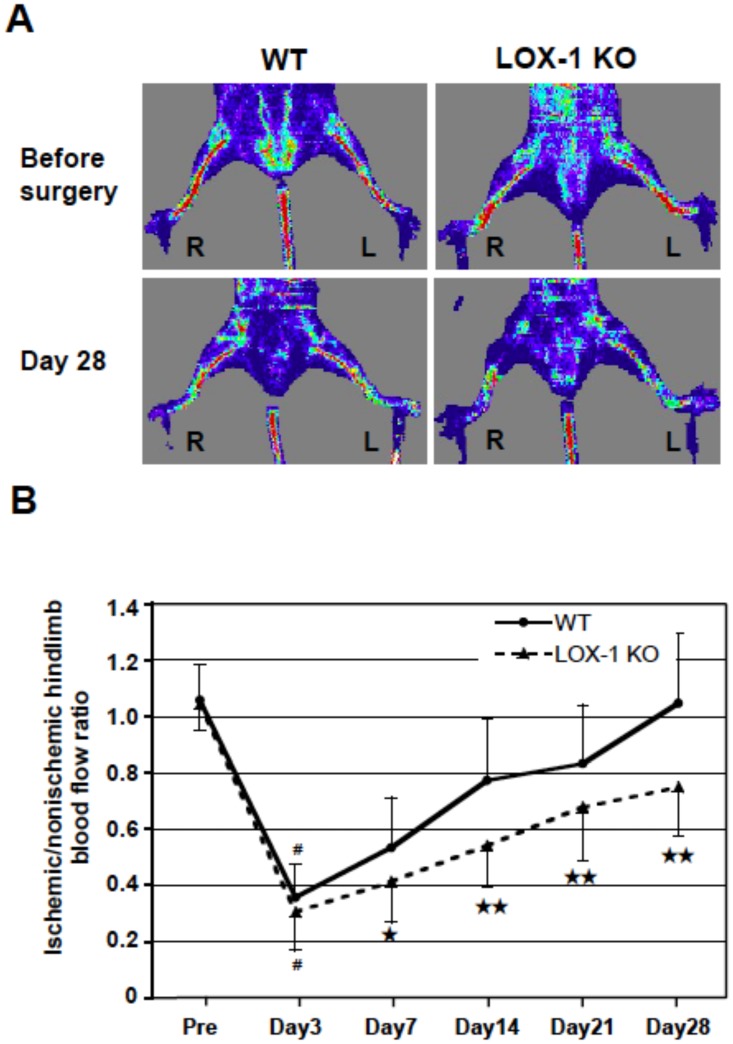
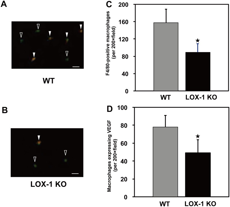
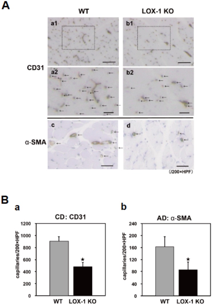
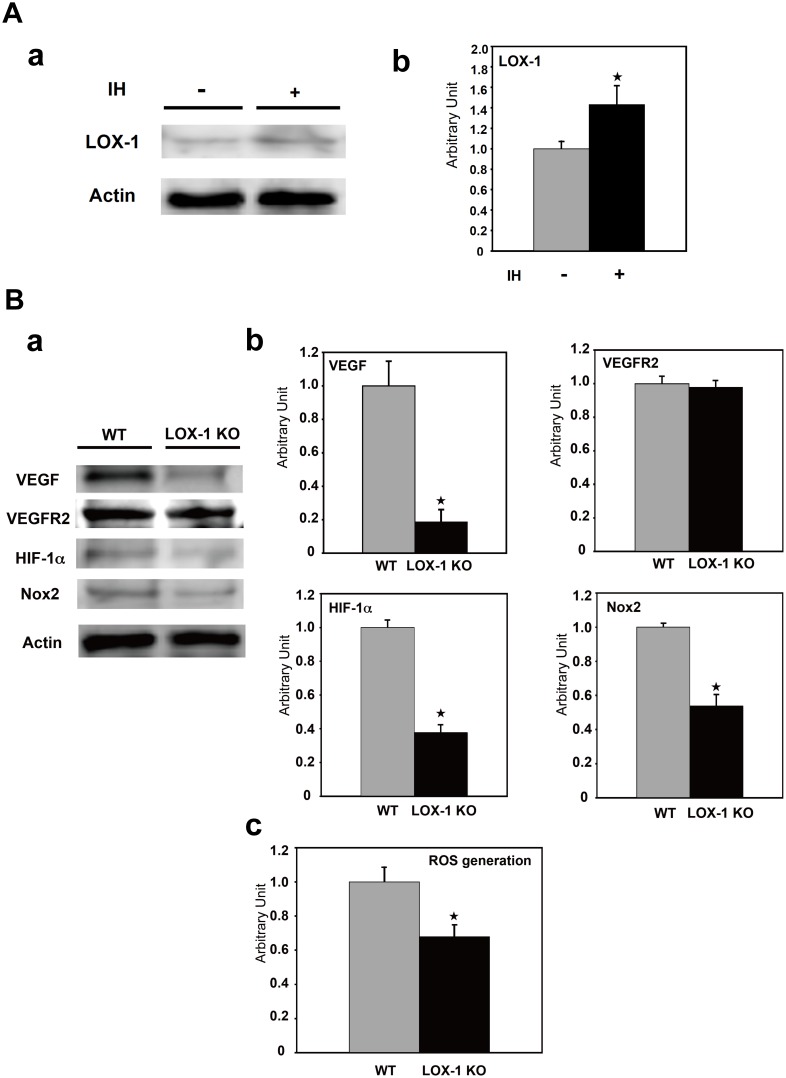
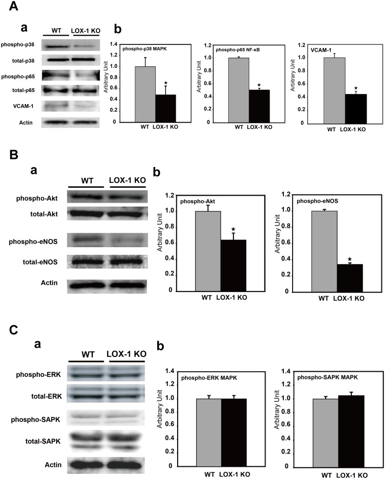
LOX-1, lectin-like oxidized low-density lipoprotein (LDL) receptor-1, is a single transmembrane receptor mainly expressed on endothelial cells. LOX-1 mediates the uptake of oxidized LDL, an early step in atherosclerosis; however, little is known about whether LOX-1 is involved in angiogenesis during tissue ischemia. Therefore, we examined the role of LOX-1 in ischemia-induced angiogenesis in the hindlimbs of LOX-1 knockout (KO) mice. Angiogenesis was evaluated in a surgically induced hindlimb ischemia model using laser Doppler blood flowmetry (LDBF) and histological capillary density (CD) and arteriole density (AD). After right hindlimb ischemia, the ischemic/nonischemic hindlimb blood flow ratio was persistently lower in LOX-1 KO mice than in wild-type (WT) mice. CD and AD were significantly smaller in LOX-1 KO mice than in WT mice on postoperative day 14. Immunohistochemical analysis revealed that the number of macrophages infiltrating ischemic tissues was significantly smaller in LOX-1 KO mice than in WT mice. The number of infiltrated macrophages expressing VEGF was also significantly smaller in LOX-1 KO mice than in WT mice. Western blot analysis and ROS production assay revealed that LOX- KO mice show significant decrease in Nox2 expression, ROS production and HIF-1α expression, the phosphorylation of p38 MAPK and NF-κB p65 subunit as well as expression of redox-sensitive vascular cell adhesion molecule-1 (VCAM-1) and LOX-1 itself in ischemic muscles, which is supposed to be required for macrophage infiltration expressing angiogenic factor VEGF. Reduction of VEGF expression successively suppressed the phosphorylation of Akt and eNOS, which accelerated angiogenesis, in the ischemic leg of LOX-1 KO mice. Our findings indicate that LOX-1 plays an important role in ischemia-induced angiogenesis by 1) Nox2-ROS-NF-κB activation, 2) upregulated expression of adhesion molecules: VCAM-1 and LOX-1 and 3) promoting macrophage infiltration, which expresses angiogenic factor VEGF.
凝集素样氧化低密度脂蛋白受体1(LOX-1)是一种主要在内皮细胞上表达的单跨膜受体。LOX-1介导氧化型低密度脂蛋白的摄取,这是动脉粥样硬化的早期步骤;然而,关于LOX-1在组织缺血期间是否参与血管生成知之甚少。因此,我们研究了LOX-1在LOX-1基因敲除(KO)小鼠后肢缺血诱导的血管生成中的作用。使用激光多普勒血流仪(LDBF)以及组织学毛细血管密度(CD)和小动脉密度(AD),在手术诱导的后肢缺血模型中评估血管生成。右后肢缺血后,LOX-1基因敲除小鼠的缺血/非缺血后肢血流比持续低于野生型(WT)小鼠。术后第14天,LOX-1基因敲除小鼠的CD和AD明显小于WT小鼠。免疫组织化学分析显示,LOX-1基因敲除小鼠缺血组织中浸润的巨噬细胞数量明显少于WT小鼠。表达血管内皮生长因子(VEGF)的浸润巨噬细胞数量在LOX-1基因敲除小鼠中也明显少于WT小鼠。蛋白质印迹分析和活性氧(ROS)产生测定显示,LOX-1基因敲除小鼠缺血肌肉中的Nox2表达、ROS产生和缺氧诱导因子-1α(HIF-1α)表达显著降低,p38丝裂原活化蛋白激酶(MAPK)和核因子κB p65亚基的磷酸化以及氧化还原敏感的血管细胞黏附分子-1(VCAM-1)和LOX-1自身的表达也显著降低,而这些被认为是表达血管生成因子VEGF的巨噬细胞浸润所必需的。LOX-1基因敲除小鼠缺血腿部VEGF表达的降低相继抑制了Akt和内皮型一氧化氮合酶(eNOS)的磷酸化,而Akt和eNOS的磷酸化加速了血管生成。我们的研究结果表明,LOX-1在缺血诱导的血管生成中起重要作用,其机制为:1)激活Nox2-ROS-核因子κB;2)上调黏附分子VCAM-1和LOX-1的表达;3)促进表达血管生成因子VEGF的巨噬细胞浸润。