• 文献检索
  • 文档翻译
  • 深度研究
  • 学术资讯
  • Suppr Zotero 插件Zotero 插件
  • 邀请有礼
  • 套餐&价格
  • 历史记录
应用&插件
Suppr Zotero 插件Zotero 插件浏览器插件Mac 客户端Windows 客户端微信小程序
定价
高级版会员购买积分包购买API积分包
服务
文献检索文档翻译深度研究API 文档MCP 服务
关于我们
关于 Suppr公司介绍联系我们用户协议隐私条款
关注我们

Suppr 超能文献

核心技术专利:CN118964589B侵权必究
粤ICP备2023148730 号-1Suppr @ 2026

文献检索

告别复杂PubMed语法,用中文像聊天一样搜索,搜遍4000万医学文献。AI智能推荐,让科研检索更轻松。

立即免费搜索

文件翻译

保留排版,准确专业,支持PDF/Word/PPT等文件格式,支持 12+语言互译。

免费翻译文档

深度研究

AI帮你快速写综述,25分钟生成高质量综述,智能提取关键信息,辅助科研写作。

立即免费体验

超声刺激可增加MC3T3-E1前成骨样细胞的增殖。

Ultrasound stimulation increases proliferation of MC3T3-E1 preosteoblast-like cells.

作者信息

Katiyar Amit, Duncan Randall L, Sarkar Kausik

机构信息

Department of Mechanical Engineering, University of Delaware, Newark, DE 19716, USA.

Department of Mechanical Engineering, University of Delaware, Newark, DE 19716, USA ; Department of Biological Sciences, University of Delaware, Newark, DE 19716, USA.

出版信息

J Ther Ultrasound. 2014 Jan 2;2:1. doi: 10.1186/2050-5736-2-1. eCollection 2014.

DOI:10.1186/2050-5736-2-1
PMID:25516803
原文链接:https://pmc.ncbi.nlm.nih.gov/articles/PMC4265984/
Abstract

BACKGROUND

Mechanical stimulation of bone increases bone mass and fracture healing, at least in part, through increases in proliferation of osteoblasts and osteoprogenitor cells. Researchers have previously performed in vitro studies of ultrasound-induced osteoblast proliferation but mostly used fixed ultrasound settings and have reported widely varying and inconclusive results. Here we critically investigated the effects of the excitation parameters of low-intensity pulsed ultrasound (LIPUS) stimulation on proliferation of MC3T3-E1 preosteoblastic cells in monolayer cultures.

METHODS

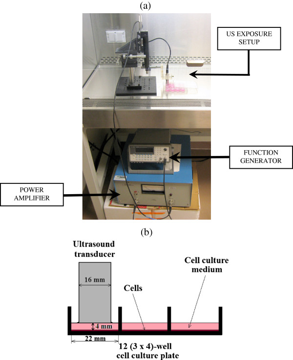
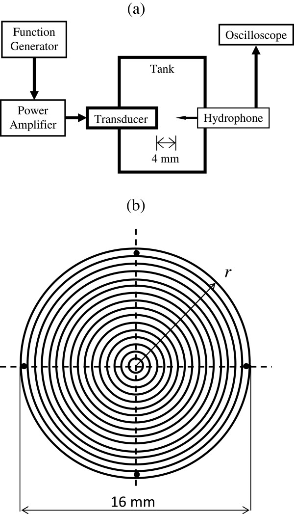
We used a custom-designed ultrasound exposure system to vary the key ultrasound parameters-intensity, frequency and excitation duration. MC3T3-E1 cells were seeded in 12-well cell culture plates. Unless otherwise specified, treated cells, in groups of three, were excited twice for 10 min with an interval of 24 h in between after cell seeding. Proliferation rates of these cells were determined using BrdU and MTS assays 24 h after the last LIPUS excitation. All data are presented as the mean ± standard error. The statistical significance was determined using Student's two-sample two-tailed t tests.

RESULTS

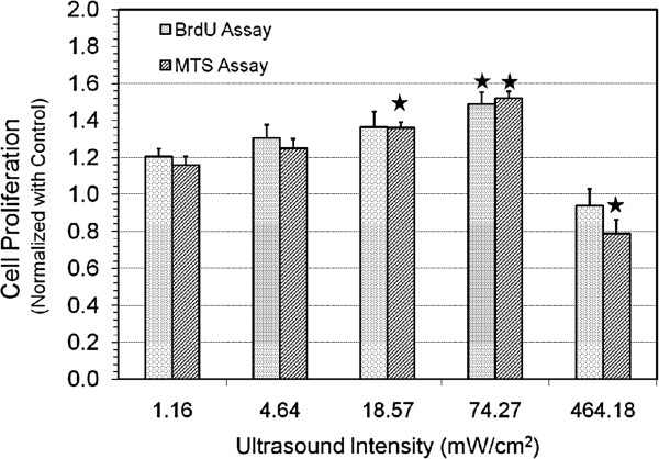
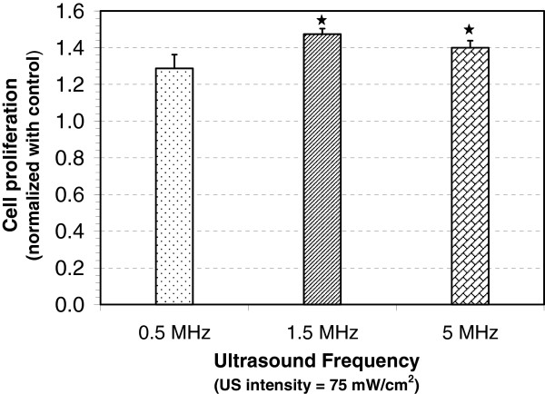
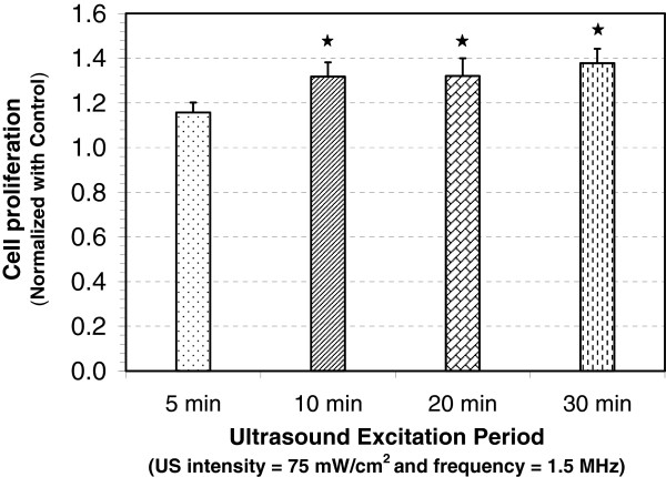
Using discrete LIPUS intensities ranging from 1 to 500 mW/cm(2) (SATA, spatial average-temporal average), we found that approximately 75 mW/cm(2) produced the greatest increase in osteoblast proliferation. Ultrasound exposures at higher intensity (approximately 465 mW/cm(2)) significantly reduced proliferation in MC3T3-E1 cells, suggesting that high-intensity pulsed ultrasound may increase apoptosis or loss of adhesion in these cells. Variation in LIPUS frequency from 0.5 MHz to 5 MHz indicated that osteoblast proliferation rate was not frequency dependent. We found no difference in the increase in proliferation rate if LIPUS was applied for 30 min/day or 10 min/day, indicating a habituation response.

CONCLUSION

This study concludes that a short-term stimulation with optimum intensity can enhance proliferation of preosteoblast-like bone cells that plays an important role in bone formation and accelerated fracture healing, also suggesting a possible therapeutic treatment for reduced bone mass.

摘要

背景

对骨骼的机械刺激可增加骨量并促进骨折愈合,至少部分是通过增加成骨细胞和骨祖细胞的增殖来实现的。研究人员此前曾进行过超声诱导成骨细胞增殖的体外研究,但大多使用固定的超声设置,且报告的结果差异很大且尚无定论。在此,我们严格研究了低强度脉冲超声(LIPUS)刺激的激发参数对单层培养的MC3T3-E1前成骨细胞增殖的影响。

方法

我们使用定制设计的超声暴露系统来改变关键的超声参数——强度、频率和激发持续时间。将MC3T3-E1细胞接种于12孔细胞培养板中。除非另有说明,每组三个处理细胞在接种后每隔24小时激发两次,每次10分钟。在最后一次LIPUS激发后24小时,使用BrdU和MTS测定法测定这些细胞的增殖率。所有数据均以平均值±标准误差表示。使用学生双样本双侧t检验确定统计学显著性。

结果

使用1至500 mW/cm²(SATA,空间平均-时间平均)的离散LIPUS强度,我们发现约75 mW/cm²可使成骨细胞增殖增加最多。更高强度(约465 mW/cm²)的超声暴露显著降低了MC3T3-E1细胞的增殖,这表明高强度脉冲超声可能会增加这些细胞的凋亡或黏附丧失。LIPUS频率在0.5 MHz至5 MHz之间变化表明成骨细胞增殖率与频率无关。我们发现,每天应用LIPUS 30分钟或10分钟,增殖率的增加没有差异,这表明存在适应性反应。

结论

本研究得出结论,以最佳强度进行短期刺激可增强前成骨样骨细胞的增殖,这在骨形成和加速骨折愈合中起重要作用,也提示了一种可能用于治疗骨量减少的方法。

https://cdn.ncbi.nlm.nih.gov/pmc/blobs/0508/4265984/e638f200b347/2050-5736-2-1-6.jpg
https://cdn.ncbi.nlm.nih.gov/pmc/blobs/0508/4265984/078069393f6b/2050-5736-2-1-1.jpg
https://cdn.ncbi.nlm.nih.gov/pmc/blobs/0508/4265984/000df37562c8/2050-5736-2-1-2.jpg
https://cdn.ncbi.nlm.nih.gov/pmc/blobs/0508/4265984/bae8c1c9f015/2050-5736-2-1-3.jpg
https://cdn.ncbi.nlm.nih.gov/pmc/blobs/0508/4265984/403add36d7e0/2050-5736-2-1-4.jpg
https://cdn.ncbi.nlm.nih.gov/pmc/blobs/0508/4265984/7f233dc70958/2050-5736-2-1-5.jpg
https://cdn.ncbi.nlm.nih.gov/pmc/blobs/0508/4265984/e638f200b347/2050-5736-2-1-6.jpg

相似文献

1
Ultrasound stimulation increases proliferation of MC3T3-E1 preosteoblast-like cells.超声刺激可增加MC3T3-E1前成骨样细胞的增殖。
J Ther Ultrasound. 2014 Jan 2;2:1. doi: 10.1186/2050-5736-2-1. eCollection 2014.
2
Intensity-related differences in collagen post-translational modification in MC3T3-E1 osteoblasts after exposure to low- and high-intensity pulsed ultrasound.暴露于低强度和高强度脉冲超声后,MC3T3-E1成骨细胞中胶原蛋白翻译后修饰的强度相关差异。
Bone. 2004 Sep;35(3):644-55. doi: 10.1016/j.bone.2004.04.024.
3
Low-intensity pulsed ultrasound enhances the positive effects of high-intensity treadmill exercise on bone in rats.低强度脉冲超声增强了高强度跑步机运动对大鼠骨的正向作用。
J Bone Miner Metab. 2023 Sep;41(5):592-605. doi: 10.1007/s00774-023-01439-6. Epub 2023 Jun 4.
4
MSTN is a key mediator for low-intensity pulsed ultrasound preventing bone loss in hindlimb-suspended rats.MSTN 是低强度脉冲超声防止后肢悬吊大鼠骨丢失的关键介质。
Bone. 2021 Feb;143:115610. doi: 10.1016/j.bone.2020.115610. Epub 2020 Aug 21.
5
Genes responsive to low-intensity pulsed ultrasound in MC3T3-E1 preosteoblast cells.MC3T3-E1 前成骨细胞中对低强度脉冲超声有反应的基因。
Int J Mol Sci. 2013 Nov 18;14(11):22721-40. doi: 10.3390/ijms141122721.
6
Study on the Effects of Low-Intensity Pulsed Ultrasound and Iron Ions for Proliferation and Differentiation of Osteoblasts.低强度脉冲超声与铁离子对成骨细胞增殖分化影响的研究。
Ultrasound Med Biol. 2024 Nov;50(11):1674-1683. doi: 10.1016/j.ultrasmedbio.2024.07.003. Epub 2024 Aug 28.
7
Low-intensity pulsed ultrasound regulates proliferation and differentiation of osteoblasts through osteocytes.低强度脉冲超声通过成骨细胞调节成骨细胞的增殖和分化。
Biochem Biophys Res Commun. 2012 Feb 10;418(2):296-300. doi: 10.1016/j.bbrc.2012.01.014. Epub 2012 Jan 13.
8
Inhibition of myostatin signal pathway may be involved in low-intensity pulsed ultrasound promoting bone healing.抑制肌生成抑制素信号通路可能参与低强度脉冲超声促进骨愈合的过程。
J Med Ultrason (2001). 2019 Oct;46(4):377-388. doi: 10.1007/s10396-019-00962-2. Epub 2019 Aug 3.
9
Application of ultrasound stimulation in bone tissue engineering.超声刺激在骨组织工程中的应用。
Int J Stem Cells. 2010 May;3(2):74-9. doi: 10.15283/ijsc.2010.3.2.74.
10
Low-intensity pulsed ultrasound-induced ATP increases bone formation via the P2X7 receptor in osteoblast-like MC3T3-E1 cells.低强度脉冲超声诱导的三磷酸腺苷通过成骨样MC3T3-E1细胞中的P2X7受体增加骨形成。
FEBS Lett. 2015 Jan 30;589(3):310-8. doi: 10.1016/j.febslet.2014.12.013. Epub 2014 Dec 24.

引用本文的文献

1
Acoustic technologies for the orchestration of cellular functions for therapeutic applications.用于编排细胞功能以实现治疗应用的声学技术。
Sci Adv. 2025 Jul 18;11(29):eadu4759. doi: 10.1126/sciadv.adu4759.
2
Piezoelectricity of hexagonal boron nitrides improves bone tissue generation as tested on osteoblasts.在成骨细胞上进行测试时,六方氮化硼的压电性可促进骨组织生成。
Beilstein J Nanotechnol. 2025 Jul 7;16:1068-1081. doi: 10.3762/bjnano.16.78. eCollection 2025.
3
Time-Dependent Effects of Low-Intensity Pulsed Ultrasound on Apoptosis and Autophagy in Malignant Melanoma Stem Cells.

本文引用的文献

1
Mechanotransduction activates α₅β₁ integrin and PI3K/Akt signaling pathways in mandibular osteoblasts.机械转导激活下颌骨成骨细胞中的 α₅β₁ 整合素和 PI3K/Akt 信号通路。
Exp Cell Res. 2011 Nov 1;317(18):2642-9. doi: 10.1016/j.yexcr.2011.07.015. Epub 2011 Jul 29.
2
Effects of combined mechanical stimulation on the proliferation and differentiation of pre-osteoblasts.联合机械刺激对前成骨细胞增殖和分化的影响。
Exp Mol Med. 2011 Jun 30;43(6):367-73. doi: 10.3858/emm.2011.43.6.040.
3
Long-term (trophic) purinergic signalling: purinoceptors control cell proliferation, differentiation and death.
低强度脉冲超声对恶性黑色素瘤干细胞凋亡和自噬的时间依赖性影响
J Cell Mol Med. 2025 Jun;29(12):e70687. doi: 10.1111/jcmm.70687.
4
A comparative study of experimental and simulated ultrasound beam propagation through cranial bones.实验与模拟超声束通过颅骨传播的对比研究。
Phys Med Biol. 2025 Jan 15;70(2):025007. doi: 10.1088/1361-6560/ada19d.
5
Microenvironment-targeted strategy steers advanced bone regeneration.微环境靶向策略引领先进的骨再生。
Mater Today Bio. 2023 Jul 21;22:100741. doi: 10.1016/j.mtbio.2023.100741. eCollection 2023 Oct.
6
Low-intensity pulsed ultrasound enhances the positive effects of high-intensity treadmill exercise on bone in rats.低强度脉冲超声增强了高强度跑步机运动对大鼠骨的正向作用。
J Bone Miner Metab. 2023 Sep;41(5):592-605. doi: 10.1007/s00774-023-01439-6. Epub 2023 Jun 4.
7
Synergistic Effect of Growth Factor Releasing Polymeric Nanoparticles and Ultrasound Stimulation on Osteogenic Differentiation.生长因子释放聚合物纳米颗粒与超声刺激对成骨分化的协同作用
Pharmaceutics. 2021 Mar 27;13(4):457. doi: 10.3390/pharmaceutics13040457.
8
Piezo1 channel activation in response to mechanobiological acoustic radiation force in osteoblastic cells.成骨细胞中Piezo1通道对机械生物学声辐射力的激活。
Bone Res. 2021 Mar 10;9(1):16. doi: 10.1038/s41413-020-00124-y.
9
Zinc Oxide Nanocrystals and High-Energy Shock Waves: A New Synergy for the Treatment of Cancer Cells.氧化锌纳米晶体与高能冲击波:治疗癌细胞的新协同作用
Front Bioeng Biotechnol. 2020 Jun 5;8:577. doi: 10.3389/fbioe.2020.00577. eCollection 2020.
10
Low-intensity pulsed ultrasound promotes the expression of immediate-early genes in mouse ST2 bone marrow stromal cells.低强度脉冲超声促进小鼠ST2骨髓基质细胞中即早基因的表达。
J Med Ultrason (2001). 2020 Apr;47(2):193-201. doi: 10.1007/s10396-020-01007-9. Epub 2020 Feb 5.
长期(营养)嘌呤能信号转导:嘌呤能受体控制细胞增殖、分化和死亡。
Cell Death Dis. 2010;1(1):e9. doi: 10.1038/cddis.2009.11.
4
Indirect low-intensity ultrasonic stimulation for tissue engineering.间接低强度超声刺激在组织工程中的应用。
J Tissue Eng. 2010 May 23;2010:973530. doi: 10.4061/2010/973530.
5
Osteogenic differentiation of rat bone marrow stromal cells by various intensities of low-intensity pulsed ultrasound.不同强度的低强度脉冲超声对大鼠骨髓基质细胞的成骨分化作用。
Ultrasonics. 2011 Apr;51(3):281-8. doi: 10.1016/j.ultras.2010.09.004. Epub 2010 Sep 26.
6
Cyclic Hydraulic Pressure and Fluid Flow Differentially Modulate Cytoskeleton Re-Organization in MC3T3 Osteoblasts.循环液压和流体流动对MC3T3成骨细胞中细胞骨架的重新组织有不同的调节作用。
Cell Mol Bioeng. 2009 Mar 1;2(1):133-143. doi: 10.1007/s12195-008-0038-2.
7
Low-intensity ultrasound increases FAK, ERK-1/2, and IRS-1 expression of intact rat bones in a noncumulative manner.低强度超声以非累积的方式增加完整大鼠骨中 FAK、ERK-1/2 和 IRS-1 的表达。
Clin Orthop Relat Res. 2010 Apr;468(4):1149-56. doi: 10.1007/s11999-009-1146-6. Epub 2009 Oct 23.
8
Daily low-intensity pulsed ultrasound-mediated osteogenic differentiation in rat osteoblasts.每日低强度脉冲超声介导大鼠成骨细胞的成骨分化。
Acta Biochim Biophys Sin (Shanghai). 2009 Feb;41(2):108-15. doi: 10.1093/abbs/gmn012.
9
Low-intensity pulsed ultrasound (LIPUS) in fresh clavicle fractures: a multi-centre double blind randomised controlled trial.低强度脉冲超声(LIPUS)治疗新鲜锁骨骨折:一项多中心双盲随机对照试验
Injury. 2008 Dec;39(12):1444-52. doi: 10.1016/j.injury.2008.04.004. Epub 2008 Jul 25.
10
Low intensity pulsed ultrasound for fracture healing: a review of the clinical evidence and the associated biological mechanism of action.低强度脉冲超声促进骨折愈合:临床证据及相关生物学作用机制综述
Ultrasonics. 2008 Aug;48(4):330-8. doi: 10.1016/j.ultras.2008.02.005. Epub 2008 Mar 27.