Ahonen Saija J, Arumilli Meharji, Seppälä Eija, Hakosalo Osmo, Kaukonen Maria K, Komáromy András M, Lohi Hannes
Department of Veterinary Biosciences and Research Programs Unit, Molecular Neurology, University of Helsinki, Helsinki, Finland; The Folkhälsan Institute of Genetics, Helsinki, Finland.
Department of Small Animal Clinical Sciences, College of Veterinary Medicine, Michigan State University, East Lansing, Michigan, United States of America; Department of Clinical Studies, School of Veterinary Medicine, University of Pennsylvania, Philadelphia, Pennsylvania, United States of America.
PLoS One. 2014 Dec 17;9(12):e114552. doi: 10.1371/journal.pone.0114552. eCollection 2014.
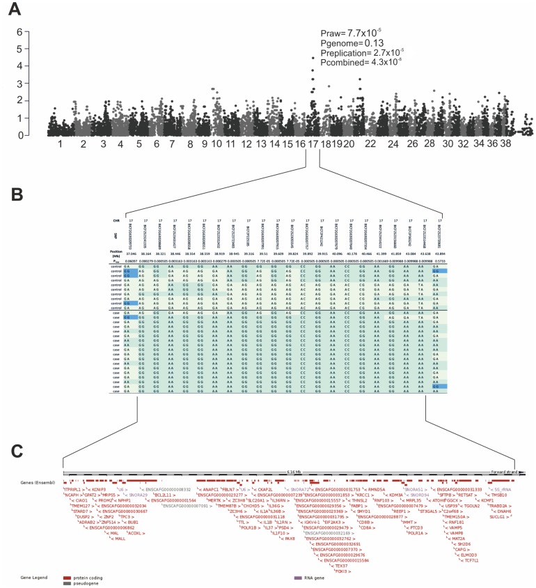
Progressive retinal degenerations are among the most common causes of blindness both in human and in dogs. Canine progressive retinal atrophy (PRA) resembles human retinitis pigmentosa (RP) and is typically characterized by a progressive loss of rod photoreceptors followed by a loss of cone function. The disease gradually progress from the loss of night and day vision to a complete blindness. We have recently described a unique form of retinopathy characterized by the multifocal gray/brown discoloration and thinning of the retina in the Swedish Vallhund (SV) breed. We aimed to identify the genetic cause by performing a genome wide association analysis in a cohort of 18 affected and 10 healthy control dogs using Illumina's canine 22k SNP array. We mapped the disease to canine chromosome 17 (p = 7.7×10(-5)) and found a 6.1 Mb shared homozygous region in the affected dogs. A combined analysis of the GWAS and replication data with additional 60 dogs confirmed the association (p = 4.3×10(-8), OR = 11.2 for homozygosity). A targeted resequencing of the entire associated region in four cases and four controls with opposite risk haplotypes identified several variants in the coding region of functional candidate genes, such as a known retinopathy gene, MERTK. However, none of the identified coding variants followed a compelling case- or breed-specific segregation pattern. The expression analyses of four candidate genes in the region, MERTK, NPHP1, ANAPC1 and KRCC1, revealed specific upregulation of MERTK in the retina of the affected dogs. Collectively, these results indicate that the retinopathy is associated with overexpression of MERTK, however further investigation is needed to discover the regulatory mutation for the better understanding of the disease pathogenesis. Our study establishes a novel gain-of-function model for the MERTK biology and provides a therapy model for retinopathy MERTK inhibitors. Meanwhile, a marker-based genetic counseling can be developed to revise breeding programs.
进行性视网膜变性是人类和犬类失明的最常见原因之一。犬类进行性视网膜萎缩(PRA)类似于人类色素性视网膜炎(RP),其典型特征是视杆光感受器逐渐丧失,随后视锥功能丧失。该疾病逐渐从丧失昼夜视力发展到完全失明。我们最近描述了一种独特的视网膜病变形式,其特征是瑞典瓦汉德犬(SV)品种的视网膜出现多灶性灰/棕色变色和变薄。我们旨在通过使用Illumina的犬类22k SNP阵列对18只患病犬和10只健康对照犬进行全基因组关联分析来确定遗传原因。我们将该疾病定位到犬类17号染色体(p = 7.7×10^(-5)),并在患病犬中发现了一个6.1 Mb的共享纯合区域。对另外60只犬的GWAS和复制数据进行联合分析证实了这种关联(p = 4.3×10^(-8),纯合性的OR = 11.2)。对具有相反风险单倍型的4例病例和4例对照的整个相关区域进行靶向重测序,在功能性候选基因的编码区域中鉴定出多个变体,例如已知的视网膜病变基因MERTK。然而,所鉴定的编码变体均未遵循令人信服的病例或品种特异性分离模式。对该区域的四个候选基因MERTK、NPHP1、ANAPC1和KRCC1进行表达分析,发现患病犬视网膜中MERTK有特异性上调。总体而言,这些结果表明视网膜病变与MERTK的过表达有关,然而需要进一步研究以发现调控突变,以便更好地理解疾病发病机制。我们的研究建立了一种新的MERTK生物学功能获得模型,并为视网膜病变提供了MERTK抑制剂治疗模型。同时,可以开发基于标记的遗传咨询来修订育种计划。