Medical Research Council Laboratory of Molecular Biology, Cambridge, UK.
Medical Research Council Laboratory of Molecular Biology, Cambridge, UK
EMBO J. 2015 Feb 3;34(3):307-25. doi: 10.15252/embj.201489847. Epub 2014 Dec 19.
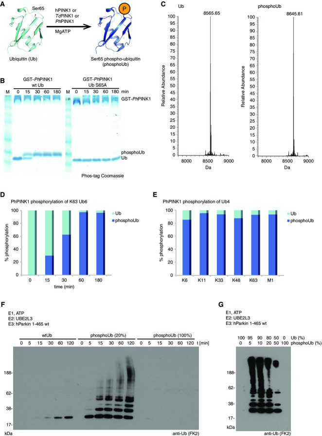
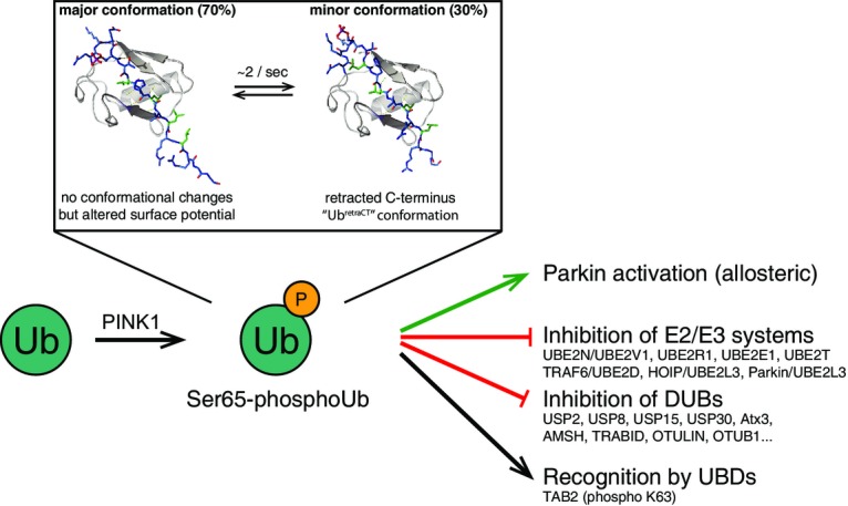
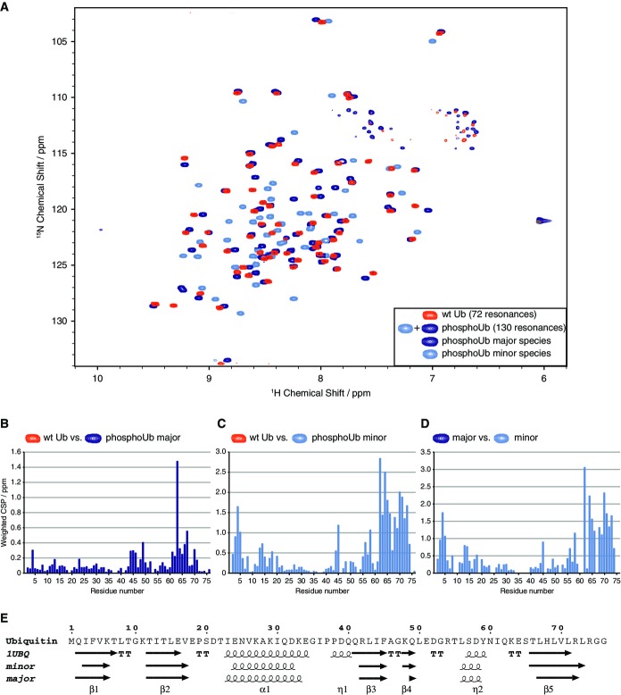
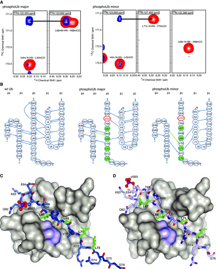
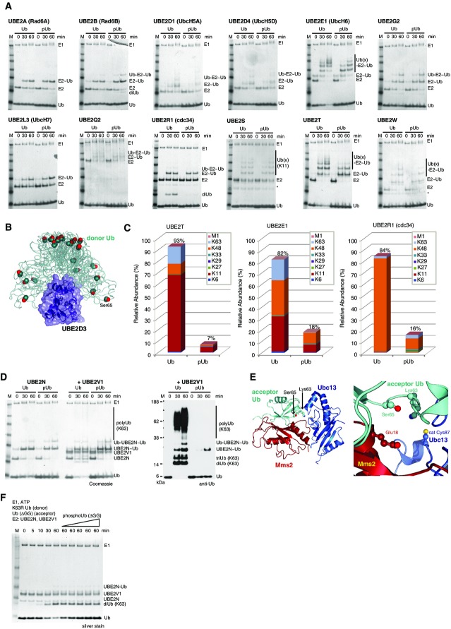
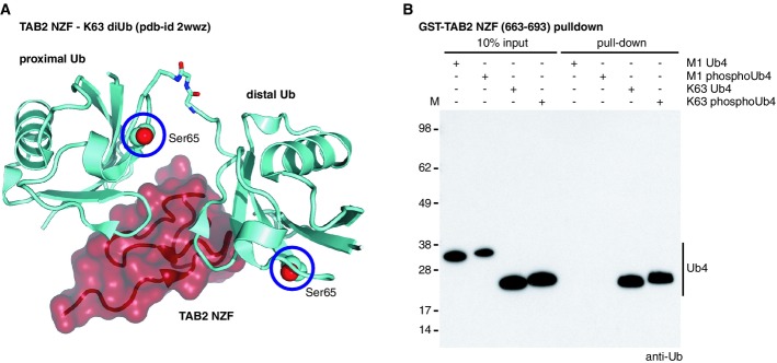
The protein kinase PINK1 was recently shown to phosphorylate ubiquitin (Ub) on Ser65, and phosphoUb activates the E3 ligase Parkin allosterically. Here, we show that PINK1 can phosphorylate every Ub in Ub chains. Moreover, Ser65 phosphorylation alters Ub structure, generating two conformations in solution. A crystal structure of the major conformation resembles Ub but has altered surface properties. NMR reveals a second phosphoUb conformation in which β5-strand slippage retracts the C-terminal tail by two residues into the Ub core. We further show that phosphoUb has no effect on E1-mediated E2 charging but can affect discharging of E2 enzymes to form polyUb chains. Notably, UBE2R1- (CDC34), UBE2N/UBE2V1- (UBC13/UEV1A), TRAF6- and HOIP-mediated chain assembly is inhibited by phosphoUb. While Lys63-linked poly-phosphoUb is recognized by the TAB2 NZF Ub binding domain (UBD), 10 out of 12 deubiquitinases (DUBs), including USP8, USP15 and USP30, are impaired in hydrolyzing phosphoUb chains. Hence, Ub phosphorylation has repercussions for ubiquitination and deubiquitination cascades beyond Parkin activation and may provide an independent layer of regulation in the Ub system.
蛋白激酶 PINK1 最近被证明可以在泛素(Ub)的丝氨酸 65 位磷酸化,磷酸化的 Ub 可以别构激活 E3 连接酶 Parkin。在这里,我们表明 PINK1 可以磷酸化 Ub 链上的每个 Ub。此外,Ser65 磷酸化改变了 Ub 的结构,在溶液中产生两种构象。主要构象的晶体结构类似于 Ub,但具有改变的表面性质。NMR 揭示了第二种磷酸化 Ub 构象,其中β5 链滑动使 C 末端尾巴向后缩回两个残基进入 Ub 核心。我们进一步表明,磷酸化 Ub 对 E1 介导的 E2 加载没有影响,但可以影响 E2 酶的释放以形成多 Ub 链。值得注意的是,UBE2R1-(CDC34)、UBE2N/UBE2V1-(UBC13/UEV1A)、TRAF6 和 HOIP 介导的链组装被磷酸化 Ub 抑制。虽然 Lys63 连接的多磷酸化 Ub 被 TAB2 NZF Ub 结合结构域(UBD)识别,但包括 USP8、USP15 和 USP30 在内的 12 种去泛素化酶(DUBs)中有 10 种在水解磷酸化 Ub 链方面受到损害。因此,Ub 磷酸化对泛素化和去泛素化级联反应的影响超出了 Parkin 的激活,并且可能在 Ub 系统中提供了一个独立的调节层。