• 文献检索
  • 文档翻译
  • 深度研究
  • 学术资讯
  • Suppr Zotero 插件Zotero 插件
  • 邀请有礼
  • 套餐&价格
  • 历史记录
应用&插件
Suppr Zotero 插件Zotero 插件浏览器插件Mac 客户端Windows 客户端微信小程序
定价
高级版会员购买积分包购买API积分包
服务
文献检索文档翻译深度研究API 文档MCP 服务
关于我们
关于 Suppr公司介绍联系我们用户协议隐私条款
关注我们

Suppr 超能文献

核心技术专利:CN118964589B侵权必究
粤ICP备2023148730 号-1Suppr @ 2026

文献检索

告别复杂PubMed语法,用中文像聊天一样搜索,搜遍4000万医学文献。AI智能推荐,让科研检索更轻松。

立即免费搜索

文件翻译

保留排版,准确专业,支持PDF/Word/PPT等文件格式,支持 12+语言互译。

免费翻译文档

深度研究

AI帮你快速写综述,25分钟生成高质量综述,智能提取关键信息,辅助科研写作。

立即免费体验

肝包膜回缩:MRI诊断范围

Hepatic capsular retraction: spectrum of diagnosis at MRI.

作者信息

Da Ines David, Mons Antoine, Braidy Chadi, Montoriol Pierre François, Garcier Jean-Marc, Vilgrain Valérie

机构信息

CHU Clermont-Ferrand, CHU Estaing, Department of Radiology and Medical Imaging, Clermont-Ferrand, France.

Assistance-Publique Hôpitaux de Paris, APHP, Hôpital Beaujon, Department of Radiology, Clichy, France ; Université Paris Diderot, Sorbonne Paris Cité, INSERM Centre de Recherche Biomédicale Bichat Beaujon, Paris, France.

出版信息

Acta Radiol Short Rep. 2014 Dec 4;3(11):2047981614545667. doi: 10.1177/2047981614545667. eCollection 2014 Dec.

DOI:10.1177/2047981614545667
PMID:25535571
原文链接:https://pmc.ncbi.nlm.nih.gov/articles/PMC4271716/
Abstract

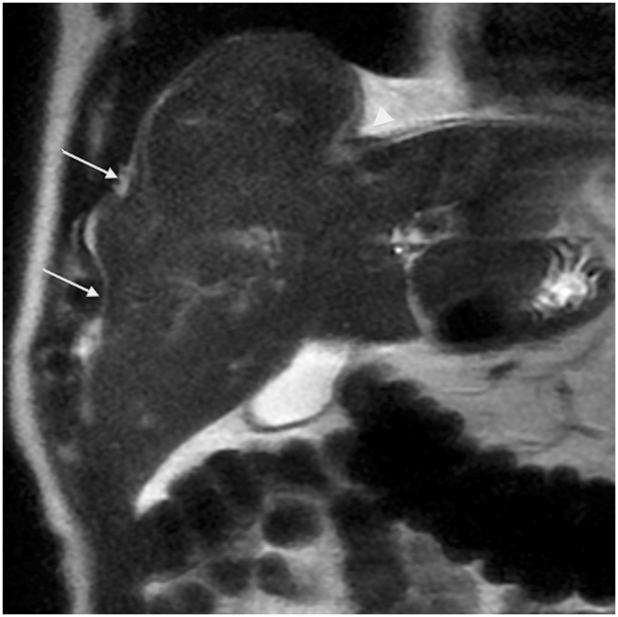
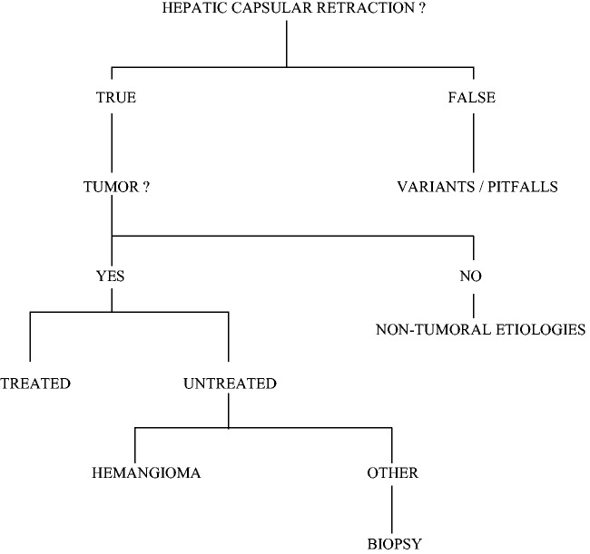
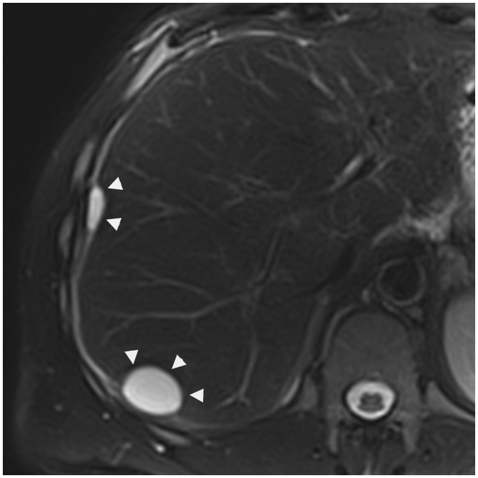
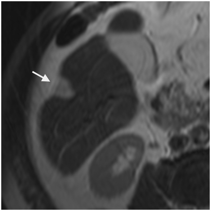
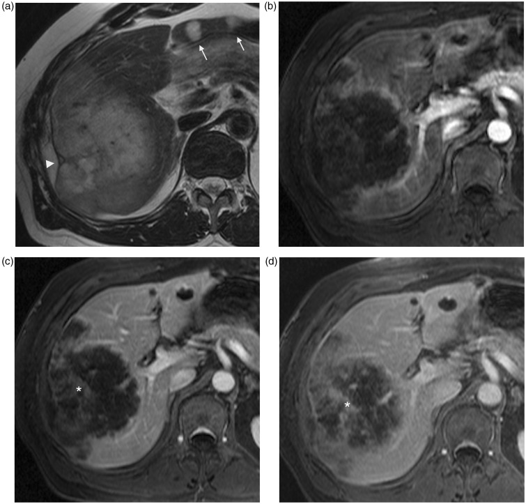
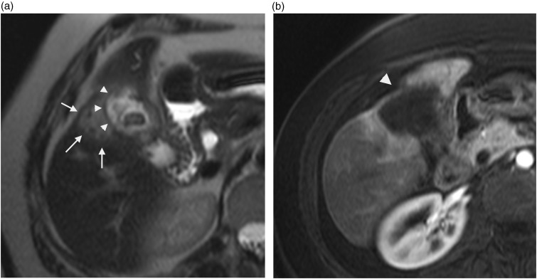
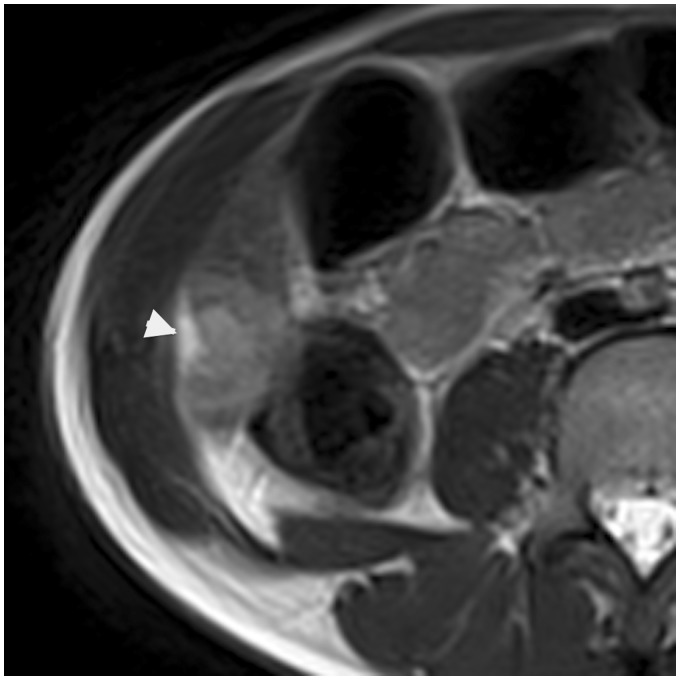
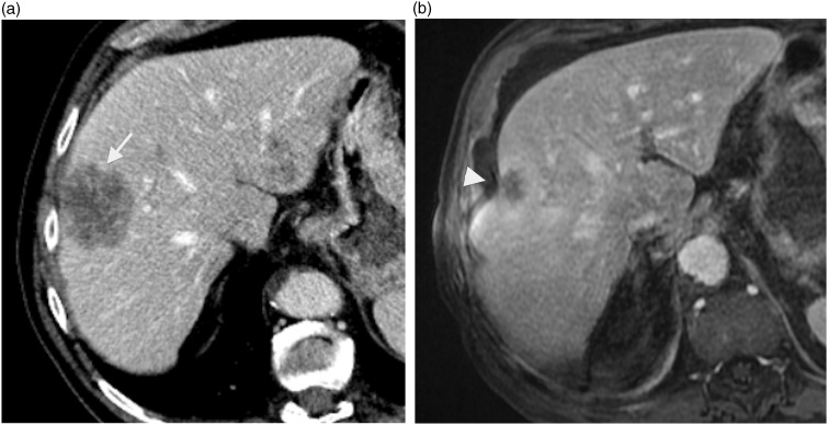
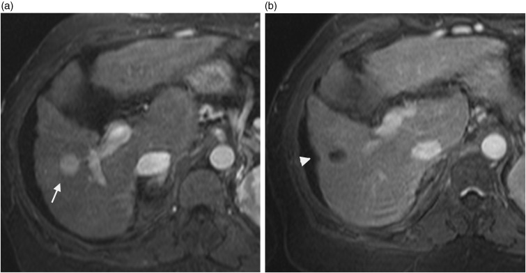
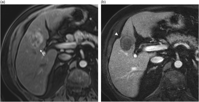
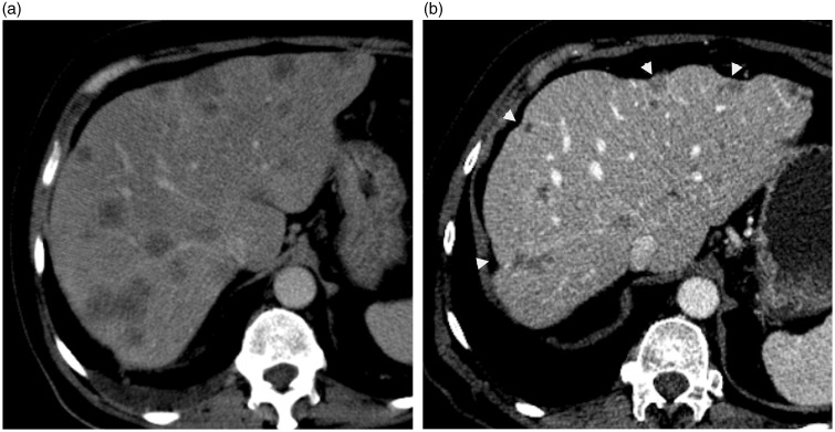
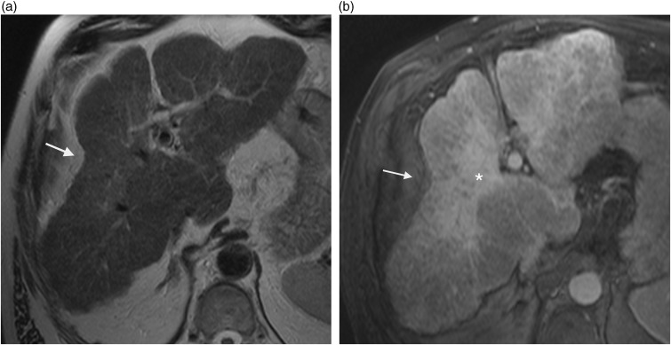
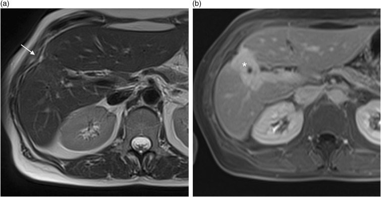
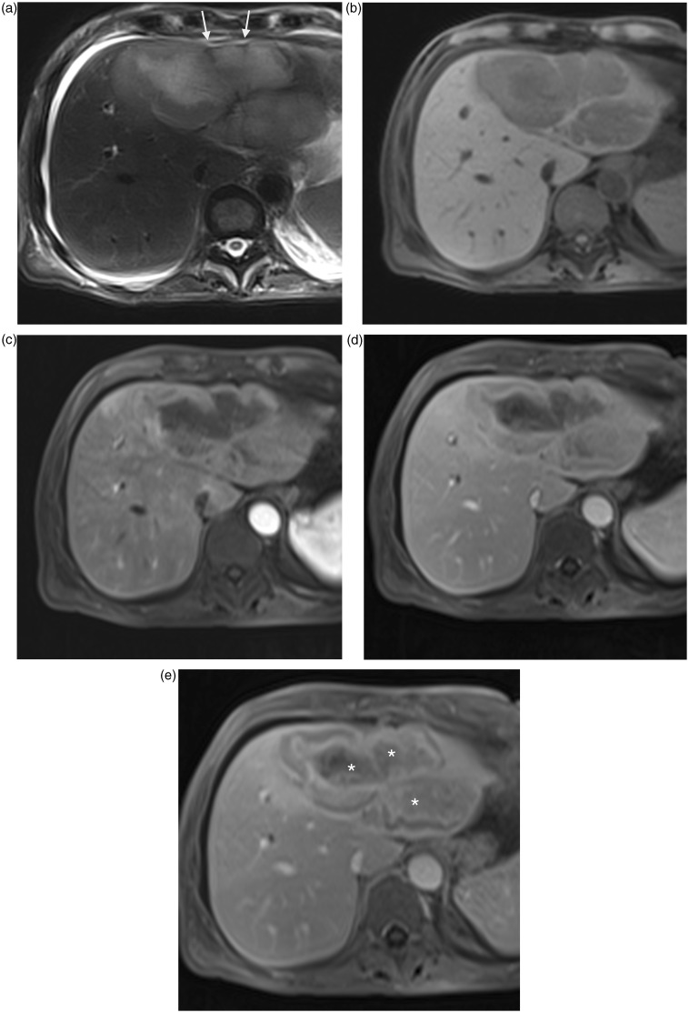
Hepatic capsular retraction is an imaging feature that deserves the attention of the radiologist. Hepatic capsular retraction is associated with a number of hepatic lesions, benign or malignant, treated or untreated. The purpose of this pictorial review is to discuss the most common benign and malignant hepatic lesions associated with this feature with an emphasis on magnetic resonance imaging (MRI).

摘要

肝包膜回缩是一种值得放射科医生关注的影像学特征。肝包膜回缩与多种肝脏病变相关,包括良性或恶性、已治疗或未治疗的病变。本图谱综述的目的是讨论与该特征相关的最常见的良性和恶性肝脏病变,重点是磁共振成像(MRI)。

https://cdn.ncbi.nlm.nih.gov/pmc/blobs/0870/4271716/ebf2ffca08a3/10.1177_2047981614545667-fig14.jpg
https://cdn.ncbi.nlm.nih.gov/pmc/blobs/0870/4271716/e5ceef787400/10.1177_2047981614545667-fig1.jpg
https://cdn.ncbi.nlm.nih.gov/pmc/blobs/0870/4271716/7181c72ede0c/10.1177_2047981614545667-fig2.jpg
https://cdn.ncbi.nlm.nih.gov/pmc/blobs/0870/4271716/edd8df2fd048/10.1177_2047981614545667-fig3.jpg
https://cdn.ncbi.nlm.nih.gov/pmc/blobs/0870/4271716/0e9f779a8a2f/10.1177_2047981614545667-fig4.jpg
https://cdn.ncbi.nlm.nih.gov/pmc/blobs/0870/4271716/b377d758f4f9/10.1177_2047981614545667-fig5.jpg
https://cdn.ncbi.nlm.nih.gov/pmc/blobs/0870/4271716/c0b0734a38d4/10.1177_2047981614545667-fig6.jpg
https://cdn.ncbi.nlm.nih.gov/pmc/blobs/0870/4271716/1a932e3c43fb/10.1177_2047981614545667-fig7.jpg
https://cdn.ncbi.nlm.nih.gov/pmc/blobs/0870/4271716/b4f5a9843fe6/10.1177_2047981614545667-fig8.jpg
https://cdn.ncbi.nlm.nih.gov/pmc/blobs/0870/4271716/0cf4b4651fc6/10.1177_2047981614545667-fig9.jpg
https://cdn.ncbi.nlm.nih.gov/pmc/blobs/0870/4271716/af993a99a3f0/10.1177_2047981614545667-fig10.jpg
https://cdn.ncbi.nlm.nih.gov/pmc/blobs/0870/4271716/ce95b5be4ca4/10.1177_2047981614545667-fig11.jpg
https://cdn.ncbi.nlm.nih.gov/pmc/blobs/0870/4271716/c29e353f9a91/10.1177_2047981614545667-fig12.jpg
https://cdn.ncbi.nlm.nih.gov/pmc/blobs/0870/4271716/38fbe7e4b3b5/10.1177_2047981614545667-fig13.jpg
https://cdn.ncbi.nlm.nih.gov/pmc/blobs/0870/4271716/ebf2ffca08a3/10.1177_2047981614545667-fig14.jpg

相似文献

1
Hepatic capsular retraction: spectrum of diagnosis at MRI.肝包膜回缩:MRI诊断范围
Acta Radiol Short Rep. 2014 Dec 4;3(11):2047981614545667. doi: 10.1177/2047981614545667. eCollection 2014 Dec.
2
[Imaging manifestations and pathologic basis for hepatic capsular retraction syndrome caused by benign and malignant liver tumors].[良恶性肝脏肿瘤所致肝包膜回缩综合征的影像学表现及病理基础]
Zhong Nan Da Xue Xue Bao Yi Xue Ban. 2015 Oct;40(10):1132-7. doi: 10.11817/j.issn.1672-7347.2015.10.013.
3
Hepatic capsular retraction: An updated MR imaging review.肝脏包膜回缩:更新的磁共振成像综述。
Eur J Radiol. 2019 Apr;113:15-23. doi: 10.1016/j.ejrad.2019.01.037. Epub 2019 Jan 31.
4
Causes of hepatic capsular retraction: a pictorial essay.肝包膜回缩的原因:图文综述
Insights Imaging. 2016 Dec;7(6):831-840. doi: 10.1007/s13244-016-0520-7. Epub 2016 Sep 29.
5
Liver lesions with hepatic capsular retraction.伴有肝包膜回缩的肝脏病变。
Semin Ultrasound CT MR. 2009 Oct;30(5):426-35. doi: 10.1053/j.sult.2009.06.002.
6
Capsular retraction of the liver in malignant tumor of the biliary tract MRI findings.胆道恶性肿瘤中肝脏包膜回缩的MRI表现。
Clin Imaging. 1994 Oct-Dec;18(4):255-7. doi: 10.1016/0899-7071(94)90003-5.
7
Is capsular retraction a specific CT sign of malignant liver tumor?肝包膜回缩是恶性肝肿瘤的特异性CT征象吗?
Eur Radiol. 1999;9(8):1543-5. doi: 10.1007/s003300050880.
8
CT of hepatic tumors: prevalence and specificity of retraction of the adjacent liver capsule.肝脏肿瘤的CT检查:邻近肝包膜回缩的发生率及特异性
AJR Am J Roentgenol. 1994 May;162(5):1119-22. doi: 10.2214/ajr.162.5.8165994.
9
CT and MRI diagnosis of hepatic epithelioid hemangioendothelioma.CT 和 MRI 诊断肝上皮样血管内皮细胞瘤。
Hepatobiliary Pancreat Dis Int. 2010 Apr;9(2):154-8.
10
Pictorial review: various causes of hepatic capsular retraction: CT and MR findings.图片综述:肝包膜回缩的各种原因:CT和MR表现
Br J Radiol. 2002 Dec;75(900):994-1002. doi: 10.1259/bjr.75.900.750994.

引用本文的文献

1
Standardizing the reporting of cholangiocarcinoma: the society of abdominal radiology disease focused panel on cholangiocarinoma lexicon.胆管癌报告的标准化:腹部放射学会胆管癌词汇疾病聚焦小组
Abdom Radiol (NY). 2025 Jul;50(7):2858-2867. doi: 10.1007/s00261-024-04769-9. Epub 2025 Jan 7.
2
Usefulness of MRI Scoring System for Differential Diagnosis between Xanthogranulomatous Cholecystitis and Wall-Thickening Type Gallbladder Cancer.MRI评分系统在黄色肉芽肿性胆囊炎与胆囊壁增厚型胆囊癌鉴别诊断中的应用价值
J Korean Soc Radiol. 2024 Jan;85(1):147-160. doi: 10.3348/jksr.2023.0036. Epub 2023 Oct 27.
3
Magnetic Resonance Imaging versus Computed Tomography for Biliary Tract Intraductal Papillary Mucinous Neoplasm (BT-IPMN): A Diagnostic Performance Analysis.

本文引用的文献

1
Clinical features, image findings, and prognosis of inflammatory pseudotumor of the liver: a multicenter experience of 45 cases.肝脏炎性假瘤的临床特征、影像学表现及预后:45 例多中心经验。
Gut Liver. 2014 Jan;8(1):58-63. doi: 10.5009/gnl.2014.8.1.58. Epub 2013 Nov 21.
2
Inflammatory pseudotumor of the liver: a case report and review of the literature.肝脏炎性假瘤:一例病例报告及文献复习
J Med Case Rep. 2011 May 21;5:196. doi: 10.1186/1752-1947-5-196.
3
Capsular retraction: an uncommon imaging finding in hepatic inflammatory pseudotumour.
磁共振成像与计算机断层扫描在胆管内乳头状黏液性肿瘤(BT-IPMN)中的诊断性能分析。
Med Sci Monit. 2020 Apr 1;26:e920952. doi: 10.12659/MSM.920952.
4
Causes of hepatic capsular retraction: a pictorial essay.肝包膜回缩的原因:图文综述
Insights Imaging. 2016 Dec;7(6):831-840. doi: 10.1007/s13244-016-0520-7. Epub 2016 Sep 29.
囊袋回缩:肝脏炎性假瘤中一种不常见的影像学表现。
Br J Radiol. 2009 Dec;82(984):e256-60. doi: 10.1259/bjr/98517258.
4
[Liver capsule retraction adjacent to a circumscribed liver lesion: review of 26 cases with histological confirmation].
J Radiol. 2009 Sep;90(9 Pt 1):1067-74. doi: 10.1016/s0221-0363(09)73246-9.
5
[Retraction of the liver capsule: a useful imaging finding].
J Radiol. 2009 Sep;90(9 Pt 1):1019-20. doi: 10.1016/s0221-0363(09)73239-1.
6
Varying appearances of cholangiocarcinoma: radiologic-pathologic correlation.胆管癌的不同表现:放射学与病理学的相关性
Radiographics. 2009 May-Jun;29(3):683-700. doi: 10.1148/rg.293085729.
7
Focal confluent fibrosis in cirrhotic liver: natural history studied with serial CT.肝硬化肝脏中的局灶性融合性纤维化:通过系列CT研究其自然史
AJR Am J Roentgenol. 2009 May;192(5):1341-7. doi: 10.2214/AJR.07.2782.
8
The effects of cancer chemotherapy on liver imaging.癌症化疗对肝脏成像的影响。
Eur Radiol. 2009 Jul;19(7):1752-62. doi: 10.1007/s00330-009-1333-6. Epub 2009 Feb 24.
9
Imaging features of sclerosed hemangioma.硬化性血管瘤的影像学特征。
AJR Am J Roentgenol. 2007 Jul;189(1):67-72. doi: 10.2214/AJR.06.1076.
10
[Inflammatory pseudo-tumor of the liver: is pre-operative diagnosis possible?].[肝脏炎性假瘤:术前能否诊断?]
Gastroenterol Clin Biol. 2003 Oct;27(10):883-90.