Jordan Paivi M, Fettis Margaret, Holt Joseph C
Department of Otolaryngology, University of Rochester, Rochester, New York.
J Comp Neurol. 2015 Jun 1;523(8):1258-80. doi: 10.1002/cne.23738. Epub 2015 Mar 25.
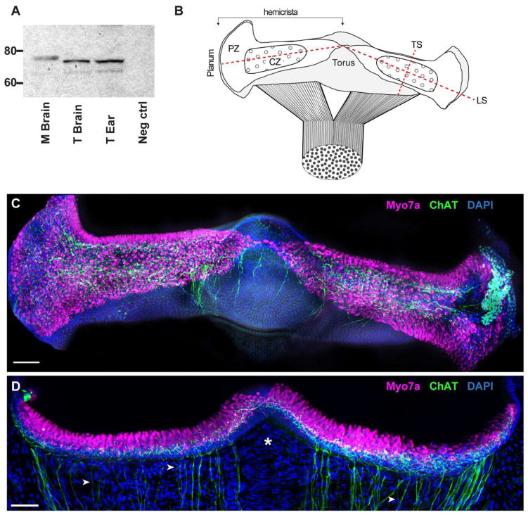
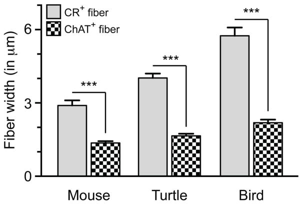
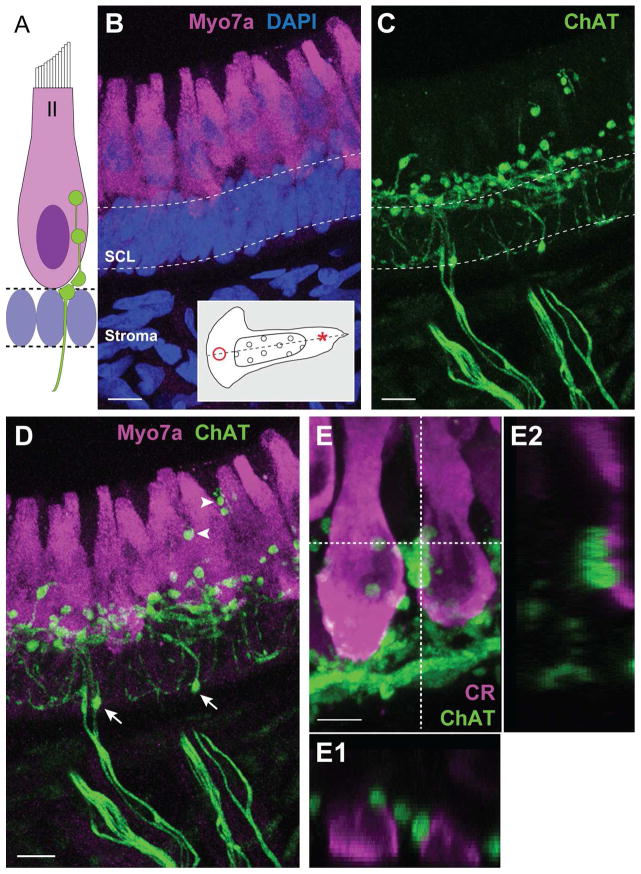
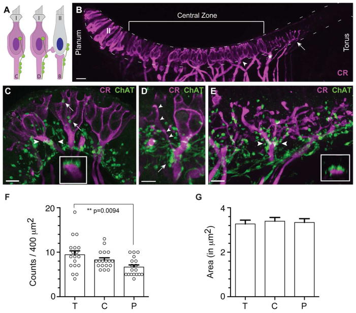
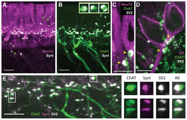
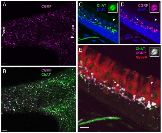
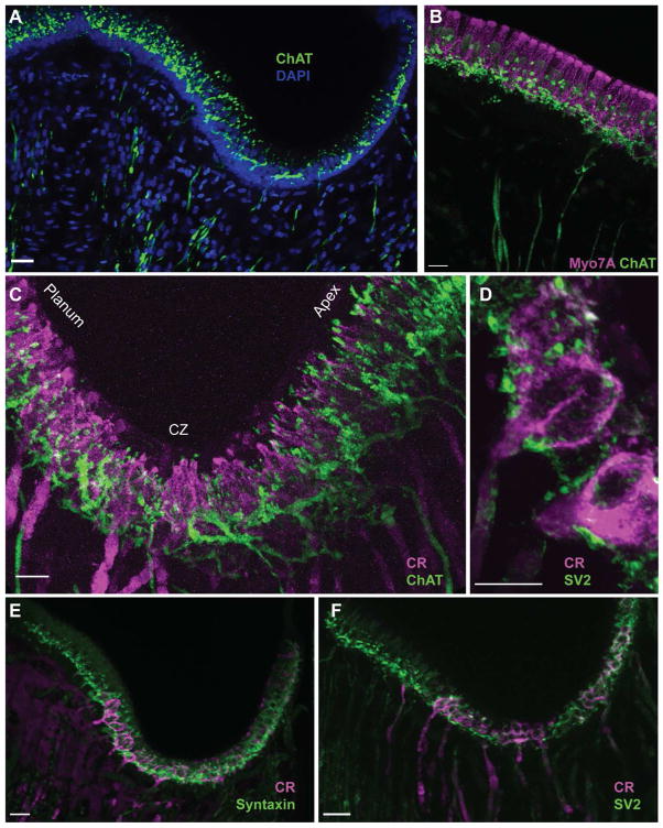
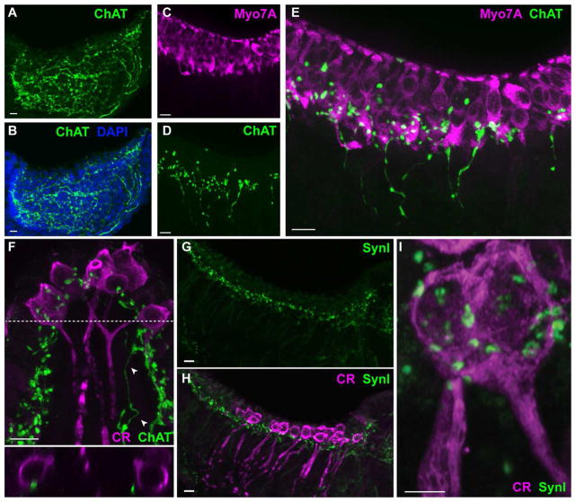
In the vestibular periphery of nearly every vertebrate, cholinergic vestibular efferent neurons give rise to numerous presynaptic varicosities that target hair cells and afferent processes in the sensory neuroepithelium. Although pharmacological studies have described the postsynaptic actions of vestibular efferent stimulation in several species, characterization of efferent innervation patterns and the relative distribution of efferent varicosities among hair cells and afferents are also integral to understanding how efferent synapses operate. Vestibular efferent markers, however, have not been well characterized in the turtle, one of the animal models used by our laboratory. Here we sought to identify reliable efferent neuronal markers in the vestibular periphery of turtle, to use these markers to understand how efferent synapses are organized, and to compare efferent neuronal labeling patterns in turtle with two other amniotes using some of the same markers. Efferent fibers and varicosities were visualized in the semicircular canal of red-eared turtles (Trachemys scripta elegans), zebra finches (Taeniopygia guttata), and mice (Mus musculus) utilizing fluorescent immunohistochemistry with antibodies against choline acetyltransferase (ChAT). Vestibular hair cells and afferents were counterstained using antibodies to myosin VIIa and calretinin. In all species, ChAT labeled a population of small diameter fibers giving rise to numerous spherical varicosities abutting type II hair cells and afferent processes. That these ChAT-positive varicosities represent presynaptic release sites were demonstrated by colabeling with antibodies against the synaptic vesicle proteins synapsin I, SV2, or syntaxin and the neuropeptide calcitonin gene-related peptide. Comparisons of efferent innervation patterns among the three species are discussed.
在几乎所有脊椎动物的前庭外周,胆碱能前庭传出神经元会产生大量突触前膨体,这些膨体靶向感觉神经上皮中的毛细胞和传入神经纤维。尽管药理学研究已经描述了几种物种中前庭传出刺激的突触后作用,但传出神经支配模式以及毛细胞和传入神经纤维之间传出膨体的相对分布特征,对于理解传出突触的运作方式同样不可或缺。然而,在我们实验室所使用的动物模型之一——乌龟中,前庭传出标记物尚未得到很好的表征。在此,我们试图在乌龟的前庭外周识别可靠的传出神经元标记物,利用这些标记物来了解传出突触是如何组织的,并使用一些相同的标记物比较乌龟与另外两种羊膜动物的传出神经元标记模式。利用针对胆碱乙酰转移酶(ChAT)的抗体进行荧光免疫组织化学,在红耳龟(滑龟)、斑胸草雀和小鼠的半规管中观察到了传出纤维和膨体。使用抗肌球蛋白VIIa和钙视网膜蛋白的抗体对前庭毛细胞和传入神经纤维进行复染。在所有物种中,ChAT标记了一群小直径纤维,这些纤维产生大量球形膨体,与II型毛细胞和传入神经纤维相邻。通过与针对突触囊泡蛋白突触素I、SV2或 syntaxin以及神经肽降钙素基因相关肽的抗体进行共标记,证明了这些ChAT阳性膨体代表突触前释放位点。讨论了这三个物种之间传出神经支配模式的比较。