Li Linwei, Mirza Shamaruh, Richardson Spencer J, Gallant Esther M, Thekkedam Chris, Pace Suzy M, Zorzato Francesco, Liu Dan, Beard Nicole A, Dulhunty Angela F
John Curtin School of Medical Research, ACT 0200, Australia.
John Curtin School of Medical Research, ACT 0200, Australia
J Cell Sci. 2015 Mar 1;128(5):951-63. doi: 10.1242/jcs.160689. Epub 2015 Jan 20.
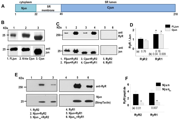
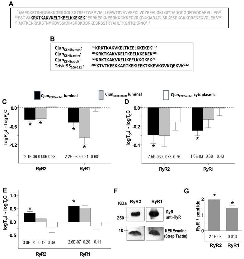
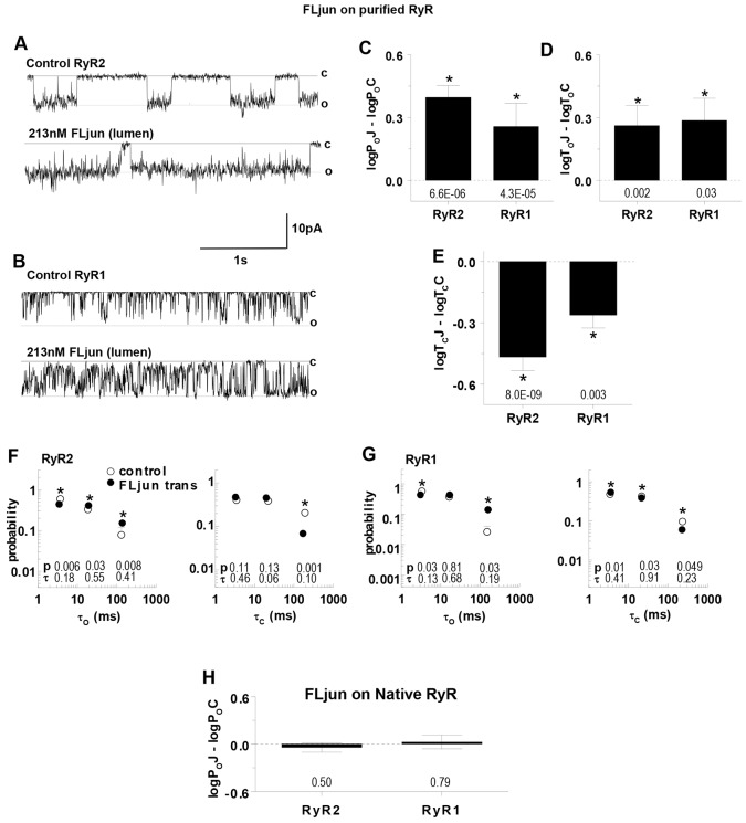
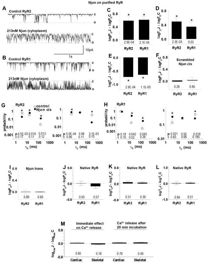
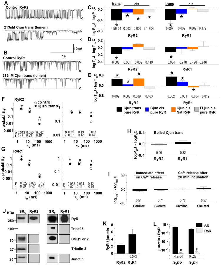
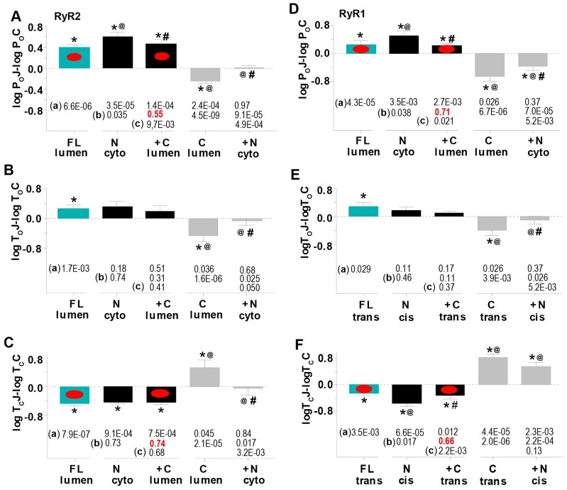
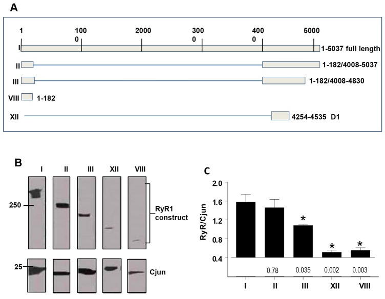
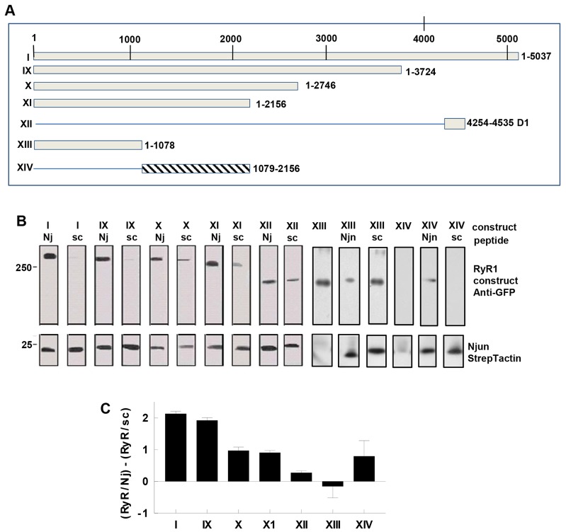
Junctin, a non-catalytic splice variant encoded by the aspartate-β-hydroxylase (Asph) gene, is inserted into the membrane of the sarcoplasmic reticulum (SR) Ca(2+) store where it modifies Ca(2+) signalling in the heart and skeletal muscle through its regulation of ryanodine receptor (RyR) Ca(2+) release channels. Junctin is required for normal muscle function as its knockout leads to abnormal Ca(2+) signalling, muscle dysfunction and cardiac arrhythmia. However, the nature of the molecular interaction between junctin and RyRs is largely unknown and was assumed to occur only in the SR lumen. We find that there is substantial binding of RyRs to full junctin, and the junctin luminal and, unexpectedly, cytoplasmic domains. Binding of these different junctin domains had distinct effects on RyR1 and RyR2 activity: full junctin in the luminal solution increased RyR channel activity by ∼threefold, the C-terminal luminal interaction inhibited RyR channel activity by ∼50%, and the N-terminal cytoplasmic binding produced an ∼fivefold increase in RyR activity. The cytoplasmic interaction between junctin and RyR is required for luminal binding to replicate the influence of full junctin on RyR1 and RyR2 activity. The C-terminal domain of junctin binds to residues including the S1-S2 linker of RyR1 and N-terminal domain of junctin binds between RyR1 residues 1078 and 2156.
连接蛋白是由天冬氨酸-β-羟化酶(Asph)基因编码的一种非催化性剪接变体,它插入到肌浆网(SR)钙库的膜中,通过调节兰尼碱受体(RyR)钙释放通道来改变心脏和骨骼肌中的钙信号。连接蛋白是正常肌肉功能所必需的,因为其基因敲除会导致异常的钙信号、肌肉功能障碍和心律失常。然而,连接蛋白与RyR之间分子相互作用的本质在很大程度上尚不清楚,并且一直被认为仅发生在肌浆网腔中。我们发现,RyR与完整的连接蛋白、连接蛋白的腔内结构域以及出乎意料的胞质结构域之间存在大量结合。这些不同的连接蛋白结构域的结合对RyR1和RyR2的活性有不同的影响:腔内溶液中的完整连接蛋白使RyR通道活性增加约三倍,C末端腔内相互作用使RyR通道活性降低约50%,而N末端胞质结合使RyR活性增加约五倍。连接蛋白与RyR之间的胞质相互作用是腔内结合复制完整连接蛋白对RyR1和RyR2活性影响所必需的。连接蛋白的C末端结构域与包括RyR1的S1-S2连接区在内的残基结合,连接蛋白的N末端结构域在RyR1的1078和2156位残基之间结合。