• 文献检索
  • 文档翻译
  • 深度研究
  • 学术资讯
  • Suppr Zotero 插件Zotero 插件
  • 邀请有礼
  • 套餐&价格
  • 历史记录
应用&插件
Suppr Zotero 插件Zotero 插件浏览器插件Mac 客户端Windows 客户端微信小程序
定价
高级版会员购买积分包购买API积分包
服务
文献检索文档翻译深度研究API 文档MCP 服务
关于我们
关于 Suppr公司介绍联系我们用户协议隐私条款
关注我们

Suppr 超能文献

核心技术专利:CN118964589B侵权必究
粤ICP备2023148730 号-1Suppr @ 2026

文献检索

告别复杂PubMed语法,用中文像聊天一样搜索,搜遍4000万医学文献。AI智能推荐,让科研检索更轻松。

立即免费搜索

文件翻译

保留排版,准确专业,支持PDF/Word/PPT等文件格式,支持 12+语言互译。

免费翻译文档

深度研究

AI帮你快速写综述,25分钟生成高质量综述,智能提取关键信息,辅助科研写作。

立即免费体验

反射式共聚焦显微镜在良性和恶性皮肤肿瘤检查中的应用。

The use of reflectance confocal microscopy for examination of benign and malignant skin tumors.

作者信息

Wielowieyska-Szybińska Dorota, Białek-Galas Kamila, Podolec Katarzyna, Wojas-Pelc Anna

机构信息

Department of Dermatology, University Hospital, Krakow, Poland. Head of the Department: Prof. Anna Wojas-Pelc MD, PhD.

Department of Dermatology, University Hospital, Krakow, Poland. Head of the Department: Prof. Anna Wojas-Pelc MD, PhD ; Department of Dermatology, Jagiellonian University Medical College, Krakow, Poland. Head of the Department: Prof. Anna Wojas-Pelc MD, PhD.

出版信息

Postepy Dermatol Alergol. 2014 Dec;31(6):380-7. doi: 10.5114/pdia.2014.40961. Epub 2014 Dec 3.

DOI:10.5114/pdia.2014.40961
PMID:25610353
原文链接:https://pmc.ncbi.nlm.nih.gov/articles/PMC4293386/
Abstract

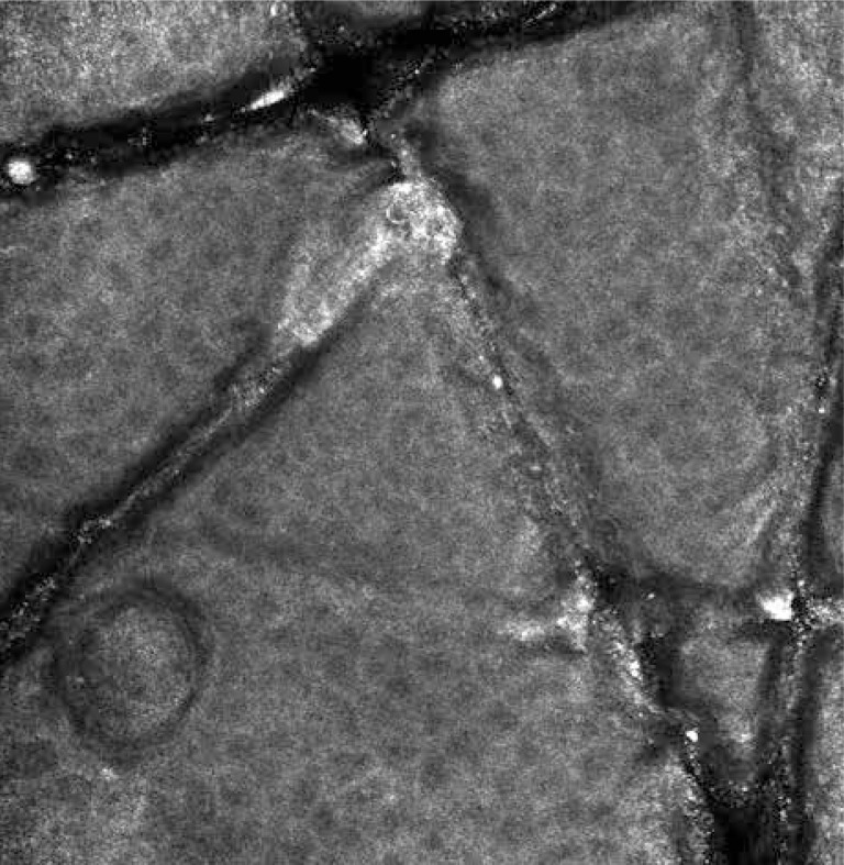
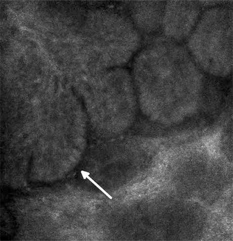
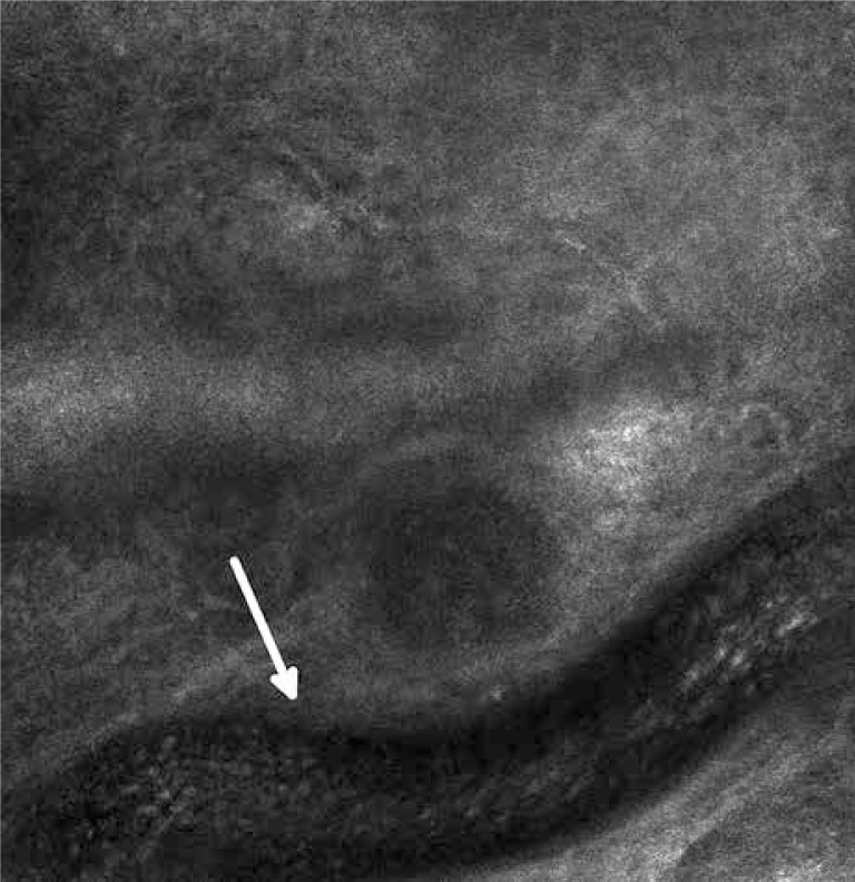
Reflectance confocal microscopy (RCM) is a modern, non-invasive diagnostic method that enables real-time imaging of epidermis and upper layers of the dermis with a nearly histological precision and high contrast. The application of this technology in skin imaging in the last few years has resulted in the progress of dermatological diagnosis, providing virtual access to the living skin erasing the need for conventional histopathology. The RCM has a potential of wide application in the dermatological diagnostic process with a particular reference to benign and malignant skin tumors. This article provides a summary of the latest reports and previous achievements in the field of RCM application in the diagnostic process of skin neoplasms. A range of dermatological indications and general characteristics of confocal images in various types of tumors are presented.

摘要

反射式共聚焦显微镜(RCM)是一种现代的非侵入性诊断方法,能够以近乎组织学的精度和高对比度对表皮和真皮上层进行实时成像。在过去几年中,这项技术在皮肤成像中的应用推动了皮肤病诊断的进展,提供了对活体皮肤的虚拟观察,不再需要传统的组织病理学检查。RCM在皮肤病诊断过程中具有广泛应用的潜力,尤其适用于良性和恶性皮肤肿瘤。本文总结了RCM在皮肤肿瘤诊断过程中的应用领域的最新报告和以往成果。介绍了一系列皮肤病学适应症以及各种肿瘤中共聚焦图像的一般特征。

https://cdn.ncbi.nlm.nih.gov/pmc/blobs/ce18/4293386/c9e4cc33f0a4/PDIA-31-22367-g003.jpg
https://cdn.ncbi.nlm.nih.gov/pmc/blobs/ce18/4293386/a2ceb613cf21/PDIA-31-22367-g001.jpg
https://cdn.ncbi.nlm.nih.gov/pmc/blobs/ce18/4293386/1e62a459acce/PDIA-31-22367-g002.jpg
https://cdn.ncbi.nlm.nih.gov/pmc/blobs/ce18/4293386/c9e4cc33f0a4/PDIA-31-22367-g003.jpg
https://cdn.ncbi.nlm.nih.gov/pmc/blobs/ce18/4293386/a2ceb613cf21/PDIA-31-22367-g001.jpg
https://cdn.ncbi.nlm.nih.gov/pmc/blobs/ce18/4293386/1e62a459acce/PDIA-31-22367-g002.jpg
https://cdn.ncbi.nlm.nih.gov/pmc/blobs/ce18/4293386/c9e4cc33f0a4/PDIA-31-22367-g003.jpg

相似文献

1
The use of reflectance confocal microscopy for examination of benign and malignant skin tumors.反射式共聚焦显微镜在良性和恶性皮肤肿瘤检查中的应用。
Postepy Dermatol Alergol. 2014 Dec;31(6):380-7. doi: 10.5114/pdia.2014.40961. Epub 2014 Dec 3.
2
The use of reflectance confocal microscopy in selected inflammatory skin diseases.反射式共聚焦显微镜在特定炎症性皮肤病中的应用。
Pol J Pathol. 2015 Jun;66(2):103-8. doi: 10.5114/pjp.2015.53001.
3
Role of In Vivo Reflectance Confocal Microscopy in the Analysis of Melanocytic Lesions.体内反射共聚焦显微镜在黑素细胞性病变分析中的作用
Acta Dermatovenerol Croat. 2018 Apr;26(1):64-67.
4
Reflectance confocal microscopy as a non-invasive diagnostic tool for Hailey-Hailey disease.反射式共聚焦显微镜作为黑利-黑利病的一种非侵入性诊断工具。
Skin Res Technol. 2014 Nov;20(4):503-9. doi: 10.1111/srt.12146. Epub 2014 Feb 17.
5
Evaluation of Syringomas by In Vivo Reflectance Confocal Microscopy: A Report of Two Cases.活体反射共聚焦显微镜评估汗管瘤:两例报告
Am J Dermatopathol. 2017 Nov;39(11):845-848. doi: 10.1097/DAD.0000000000000751.
6
Progress in the application of reflectance confocal microscopy in dermatology.反射式共聚焦显微镜在皮肤科应用中的进展。
Postepy Dermatol Alergol. 2021 Oct;38(5):709-715. doi: 10.5114/ada.2021.110077. Epub 2021 Nov 5.
7
Confocal reflectance microscopy in dermatology: promise and reality of non-invasive diagnosis and monitoring.共聚焦反射显微镜在皮肤病学中的应用:无创诊断与监测的前景与现实
Actas Dermosifiliogr. 2009 Dec;100 Suppl 2:59-69. doi: 10.1016/s0001-7310(09)73380-0.
8
Nevomelanocytic atypia detection by in vivo reflectance confocal microscopy.通过体内反射共聚焦显微镜检测痣细胞非典型性
Medicina (Kaunas). 2014;50(4):209-15. doi: 10.1016/j.medici.2014.09.008. Epub 2014 Oct 1.
9
Concordance between in vivo reflectance confocal microscopy and optical histology of lymphomatoid papulosis.蕈样肉芽肿的体内反射共聚焦显微镜与光学组织病理学的一致性。
Skin Res Technol. 2013 Aug;19(3):308-13. doi: 10.1111/srt.12046. Epub 2013 Feb 26.
10
Reflectance confocal microscopy for pigmentary disorders.反射式共聚焦显微镜在色素障碍性疾病中的应用。
Exp Dermatol. 2010 Mar;19(3):233-9. doi: 10.1111/j.1600-0625.2009.00983.x. Epub 2009 Nov 2.

引用本文的文献

1
Dermoscopic features in different dermatopathological stages of cutaneous melanomas.皮肤黑色素瘤不同皮肤病理阶段的皮肤镜特征。
Postepy Dermatol Alergol. 2020 Oct;37(5):677-684. doi: 10.5114/ada.2019.83221. Epub 2019 Mar 4.
2
Reciprocal Interplay Between Fibrillar Collagens and Collagen-Binding Integrins: Implications in Cancer Progression and Metastasis.纤维状胶原蛋白与胶原蛋白结合整合素之间的相互作用:对癌症进展和转移的影响
Front Oncol. 2020 Aug 18;10:1488. doi: 10.3389/fonc.2020.01488. eCollection 2020.
3
One Step Melanoma Surgery for Patient with Thick Primary Melanomas: "To Break the Rules, You Must First Master Them!".

本文引用的文献

1
Confocal microscopy insights into the treatment and cellular immune response of Basal cell carcinoma to photodynamic therapy.共聚焦显微镜对光动力疗法治疗基底细胞癌及其细胞免疫应答的观察。
Dermatology. 2012;225(3):264-70. doi: 10.1159/000345106. Epub 2012 Dec 13.
2
Confocal reflectance microscopy in dermatology: promise and reality of non-invasive diagnosis and monitoring.共聚焦反射显微镜在皮肤病学中的应用:无创诊断与监测的前景与现实
Actas Dermosifiliogr. 2009 Dec;100 Suppl 2:59-69. doi: 10.1016/s0001-7310(09)73380-0.
3
Use of reflectance confocal microscopy to monitor response of lentigo maligna to nonsurgical treatment.
针对原发性厚黑色素瘤患者的一步式黑色素瘤手术:“要打破规则,你必须先掌握它们!”
Open Access Maced J Med Sci. 2018 Feb 9;6(2):367-371. doi: 10.3889/oamjms.2018.084. eCollection 2018 Feb 15.
4
Optical detection and monitoring of pigmented skin lesions.色素沉着性皮肤病变的光学检测与监测
Biomed Opt Express. 2017 May 11;8(6):2946-2964. doi: 10.1364/BOE.8.002946. eCollection 2017 Jun 1.
使用反射式共聚焦显微镜监测恶性雀斑对非手术治疗的反应。
Dermatol Surg. 2010 Feb;36(2):177-84. doi: 10.1111/j.1524-4725.2009.01421.x. Epub 2009 Dec 18.
4
Determination of the refractive index of highly scattering human tissue by optical coherence tomography.通过光学相干断层扫描测定高散射人体组织的折射率
Opt Lett. 1995 Nov 1;20(21):2258. doi: 10.1364/ol.20.002258.
5
Reflectance confocal microscopy--state-of-art and research overview.反射式共聚焦显微镜——技术现状与研究综述。
Semin Cutan Med Surg. 2009 Sep;28(3):172-9. doi: 10.1016/j.sder.2009.06.004.
6
Reflectance confocal microscopy criteria for squamous cell carcinomas and actinic keratoses.鳞状细胞癌和光化性角化病的反射式共聚焦显微镜检查标准。
Arch Dermatol. 2009 Jul;145(7):766-72. doi: 10.1001/archdermatol.2009.134.
7
Development of a two-step method for the diagnosis of melanoma by reflectance confocal microscopy.通过反射式共聚焦显微镜开发一种用于黑色素瘤诊断的两步法。
J Am Acad Dermatol. 2009 Aug;61(2):216-29. doi: 10.1016/j.jaad.2009.02.014. Epub 2009 Apr 29.
8
Spitz nevi: In vivo confocal microscopic features, dermatoscopic aspects, histopathologic correlates, and diagnostic significance.斯皮茨痣:体内共聚焦显微镜特征、皮肤镜表现、组织病理学相关性及诊断意义。
J Am Acad Dermatol. 2009 Feb;60(2):236-47. doi: 10.1016/j.jaad.2008.07.061. Epub 2008 Dec 16.
9
Remodeling of the dermoepidermal junction in superficial spreading melanoma: insights gained from correlation of dermoscopy, reflectance confocal microscopy, and histopathologic analysis.浅表扩散型黑色素瘤中真皮表皮交界处的重塑:通过皮肤镜检查、反射式共聚焦显微镜检查和组织病理学分析的相关性获得的见解
Arch Dermatol. 2008 Dec;144(12):1644-9. doi: 10.1001/archdermatol.2008.504.
10
In vivo microscopic features of nodular melanomas: dermoscopy, confocal microscopy, and histopathologic correlates.结节性黑色素瘤的体内微观特征:皮肤镜检查、共聚焦显微镜检查及组织病理学相关性
Arch Dermatol. 2008 Oct;144(10):1311-20. doi: 10.1001/archderm.144.10.1311.