• 文献检索
  • 文档翻译
  • 深度研究
  • 学术资讯
  • Suppr Zotero 插件Zotero 插件
  • 邀请有礼
  • 套餐&价格
  • 历史记录
应用&插件
Suppr Zotero 插件Zotero 插件浏览器插件Mac 客户端Windows 客户端微信小程序
定价
高级版会员购买积分包购买API积分包
服务
文献检索文档翻译深度研究API 文档MCP 服务
关于我们
关于 Suppr公司介绍联系我们用户协议隐私条款
关注我们

Suppr 超能文献

核心技术专利:CN118964589B侵权必究
粤ICP备2023148730 号-1Suppr @ 2026

文献检索

告别复杂PubMed语法,用中文像聊天一样搜索,搜遍4000万医学文献。AI智能推荐,让科研检索更轻松。

立即免费搜索

文件翻译

保留排版,准确专业,支持PDF/Word/PPT等文件格式,支持 12+语言互译。

免费翻译文档

深度研究

AI帮你快速写综述,25分钟生成高质量综述,智能提取关键信息,辅助科研写作。

立即免费体验

一种使用2,2'-硫代二乙醇的快速光学透明方案用于固定小鼠脑的显微镜观察。

A rapid optical clearing protocol using 2,2'-thiodiethanol for microscopic observation of fixed mouse brain.

作者信息

Aoyagi Yuka, Kawakami Ryosuke, Osanai Hisayuki, Hibi Terumasa, Nemoto Tomomi

机构信息

Research Institute for Electronic Science, Hokkaido University, Sapporo, Hokkaido, Japan; Graduate School of Information Science and Technology, Hokkaido University, Sapporo, Hokkaido, Japan.

Research Institute for Electronic Science, Hokkaido University, Sapporo, Hokkaido, Japan; Graduate School of Information Science and Technology, Hokkaido University, Sapporo, Hokkaido, Japan; Core Research for Evolutional Science and Technology (CREST), Japan Science and Technology Agency (JST), Kawaguchi, Saitama, Japan.

出版信息

PLoS One. 2015 Jan 29;10(1):e0116280. doi: 10.1371/journal.pone.0116280. eCollection 2015.

DOI:10.1371/journal.pone.0116280
PMID:25633541
原文链接:https://pmc.ncbi.nlm.nih.gov/articles/PMC4310605/
Abstract

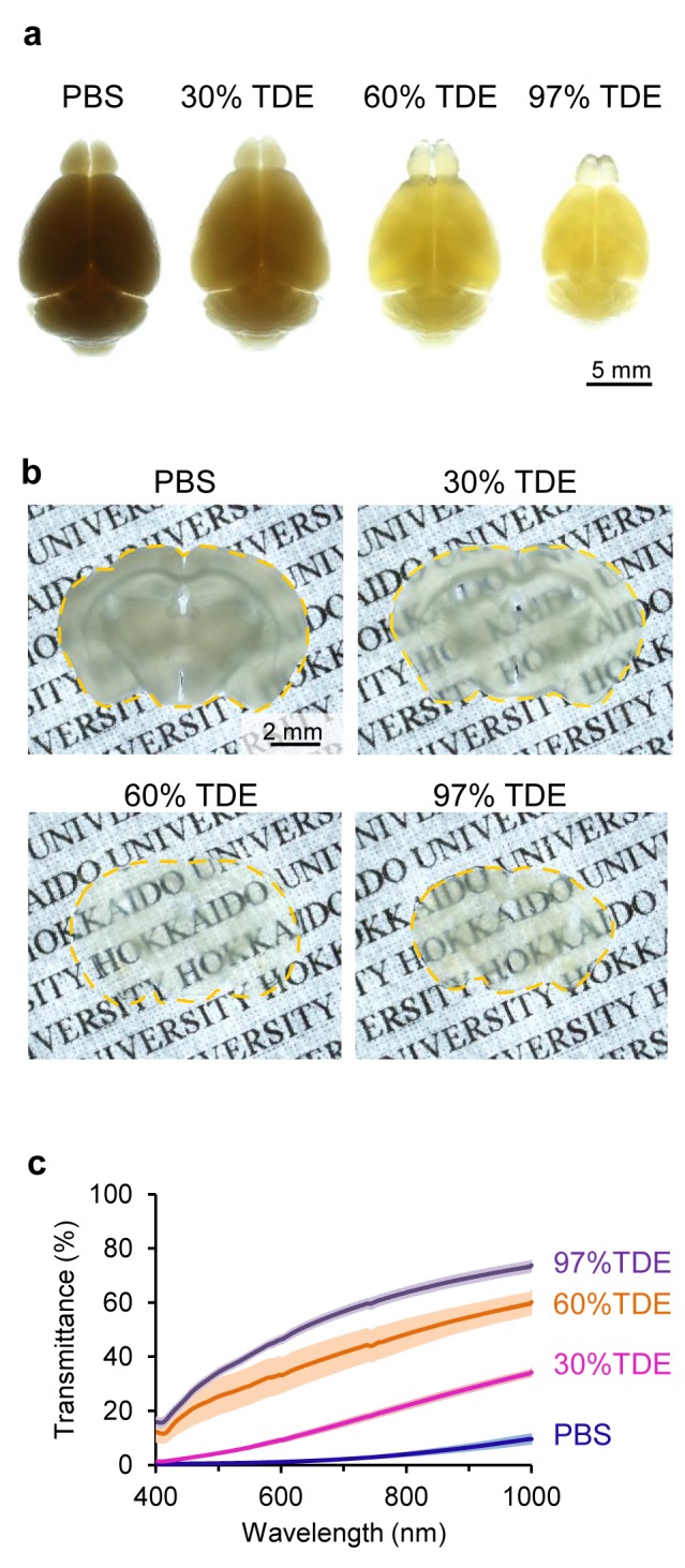
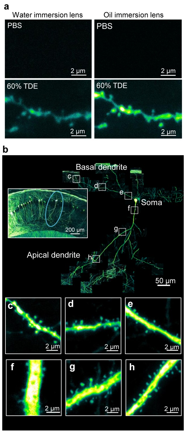
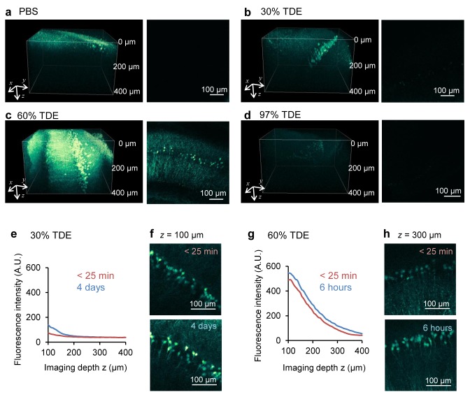
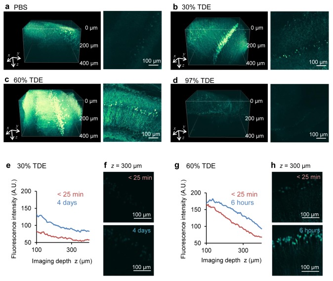
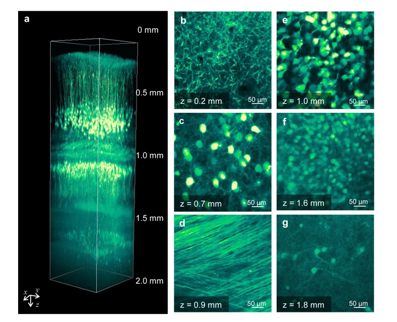
Elucidation of neural circuit functions requires visualization of the fine structure of neurons in the inner regions of thick brain specimens. However, the tissue penetration depth of laser scanning microscopy is limited by light scattering and/or absorption by the tissue. Recently, several optical clearing reagents have been proposed for visualization in fixed specimens. However, they require complicated protocols or long treatment times. Here we report the effects of 2,2'-thiodiethanol (TDE) solutions as an optical clearing reagent for fixed mouse brains expressing a yellow fluorescent protein. Immersion of fixed brains in TDE solutions rapidly (within 30 min in the case of 400-µm-thick fixed brain slices) increased their transparency and enhanced the penetration depth in both confocal and two-photon microscopy. In addition, we succeeded in visualizing dendritic spines along single dendrites at deep positions in fixed thick brain slices. These results suggest that our proposed protocol using TDE solution is a rapid and useful method for optical clearing of fixed specimens expressing fluorescent proteins.

摘要

阐明神经回路功能需要可视化厚脑标本内部区域神经元的精细结构。然而,激光扫描显微镜的组织穿透深度受组织光散射和/或吸收的限制。最近,已提出几种光学透明剂用于固定标本的可视化。然而,它们需要复杂的方案或较长的处理时间。在此,我们报告了2,2'-硫代二乙醇(TDE)溶液作为表达黄色荧光蛋白的固定小鼠脑的光学透明剂的效果。将固定脑浸入TDE溶液中可迅速(对于400μm厚的固定脑切片,在30分钟内)提高其透明度,并增强共聚焦显微镜和双光子显微镜下的穿透深度。此外,我们成功地在固定厚脑切片的深部位置沿单个树突可视化树突棘。这些结果表明,我们提出的使用TDE溶液的方案是一种快速且有用的方法,用于对表达荧光蛋白的固定标本进行光学透明处理。

https://cdn.ncbi.nlm.nih.gov/pmc/blobs/f453/4310605/a6e99771be23/pone.0116280.g005.jpg
https://cdn.ncbi.nlm.nih.gov/pmc/blobs/f453/4310605/91ef49a61008/pone.0116280.g001.jpg
https://cdn.ncbi.nlm.nih.gov/pmc/blobs/f453/4310605/0817739cb65a/pone.0116280.g002.jpg
https://cdn.ncbi.nlm.nih.gov/pmc/blobs/f453/4310605/c98971c55566/pone.0116280.g003.jpg
https://cdn.ncbi.nlm.nih.gov/pmc/blobs/f453/4310605/0ffc01ef02de/pone.0116280.g004.jpg
https://cdn.ncbi.nlm.nih.gov/pmc/blobs/f453/4310605/a6e99771be23/pone.0116280.g005.jpg

相似文献

1
A rapid optical clearing protocol using 2,2'-thiodiethanol for microscopic observation of fixed mouse brain.一种使用2,2'-硫代二乙醇的快速光学透明方案用于固定小鼠脑的显微镜观察。
PLoS One. 2015 Jan 29;10(1):e0116280. doi: 10.1371/journal.pone.0116280. eCollection 2015.
2
A Tissue Clearing Method for Neuronal Imaging from Mesoscopic to Microscopic Scales.一种用于从中尺度到微观尺度神经元成像的组织透明化方法。
J Vis Exp. 2022 May 10(183). doi: 10.3791/63941.
3
A Versatile Optical Clearing Protocol for Deep Tissue Imaging of Fluorescent Proteins in Arabidopsis thaliana.一种用于拟南芥中荧光蛋白深层组织成像的通用光学透明协议。
PLoS One. 2016 Aug 12;11(8):e0161107. doi: 10.1371/journal.pone.0161107. eCollection 2016.
4
Single-Step Fast Tissue Clearing of Thick Mouse Brain Tissue for Multi-Dimensional High-Resolution Imaging.一步法快速透明化厚鼠脑组织以进行多维高分辨率成像。
Int J Mol Sci. 2022 Jun 19;23(12):6826. doi: 10.3390/ijms23126826.
5
An advanced optical clearing protocol allows label-free detection of tissue necrosis multiphoton microscopy in injured whole muscle.一种先进的光学透明化方案,使得在损伤的整块肌肉中,利用多光子显微镜对组织坏死进行免标记检测成为可能。
Theranostics. 2021 Jan 1;11(6):2876-2891. doi: 10.7150/thno.51558. eCollection 2021.
6
A method for ultrafast tissue clearing that preserves fluorescence for multimodal and longitudinal brain imaging.一种超快组织透明化方法,可保持荧光用于多模态和纵向脑成像。
BMC Biol. 2022 Mar 29;20(1):77. doi: 10.1186/s12915-022-01275-6.
7
Super-resolution structural analysis of dendritic spines using three-dimensional structured illumination microscopy in cleared mouse brain slices.使用三维结构光照明显微镜在清除的小鼠脑片中对树突棘进行超分辨率结构分析。
Eur J Neurosci. 2018 May;47(9):1033-1042. doi: 10.1111/ejn.13901. Epub 2018 Mar 26.
8
Labeling of dendritic spines with the carbocyanine dye DiI for confocal microscopic imaging in lightly fixed cortical slices.使用碳菁染料DiI对轻度固定的皮质切片中的树突棘进行标记,用于共聚焦显微镜成像。
J Neurosci Methods. 2007 May 15;162(1-2):237-43. doi: 10.1016/j.jneumeth.2007.01.016. Epub 2007 Jan 30.
9
Comparison of Transparency and Shrinkage During Clearing of Insect Brains Using Media With Tunable Refractive Index.使用具有可调折射率的介质对昆虫大脑进行透明化处理期间的透明度和收缩率比较
Front Neuroanat. 2020 Nov 20;14:599282. doi: 10.3389/fnana.2020.599282. eCollection 2020.
10
Optical histology: a method to visualize microvasculature in thick tissue sections of mouse brain.光学组织学:一种可视化小鼠脑厚组织切片中微血管的方法。
PLoS One. 2013;8(1):e53753. doi: 10.1371/journal.pone.0053753. Epub 2013 Jan 23.

引用本文的文献

1
Advances in tissue optical clearing for 3D imaging in large animal.大型动物三维成像的组织光学透明技术进展
Front Optoelectron. 2025 Aug 18;18(1):18. doi: 10.1007/s12200-025-00162-6.
2
Practical considerations for birefringence microscopy of myelin structure: Microscope design and tissue processing for effective imaging.髓鞘结构双折射显微镜的实际考量:有效成像的显微镜设计与组织处理
Imaging Neurosci (Camb). 2024;2. doi: 10.1162/imag_a_00186. Epub 2024 May 17.
3
3D visualization of uterus and ovary: tissue clearing techniques and biomedical applications.

本文引用的文献

1
Single-cell phenotyping within transparent intact tissue through whole-body clearing.通过全身透明化在完整透明组织内进行单细胞表型分析。
Cell. 2014 Aug 14;158(4):945-958. doi: 10.1016/j.cell.2014.07.017. Epub 2014 Jul 31.
2
Whole-brain imaging with single-cell resolution using chemical cocktails and computational analysis.使用化学鸡尾酒和计算分析进行单细胞分辨率的全脑成像。
Cell. 2014 Apr 24;157(3):726-39. doi: 10.1016/j.cell.2014.03.042. Epub 2014 Apr 17.
3
Comprehensive evaluation of peripheral nerve regeneration in the acute healing phase using tissue clearing and optical microscopy in a rodent model.
子宫和卵巢的三维可视化:组织透明化技术及生物医学应用
Front Bioeng Biotechnol. 2025 Jul 7;13:1610539. doi: 10.3389/fbioe.2025.1610539. eCollection 2025.
4
Optomechanical Properties of Swine Skin Tissue Treated With a Nontoxic Optical Clearing Agent.用无毒光学透明剂处理的猪皮肤组织的光机械性能
J Biophotonics. 2025 Aug;18(8):e70007. doi: 10.1002/jbio.70007. Epub 2025 Mar 13.
5
Visualization of porcine and human aqueous humor outflow tract anatomies with transparency enhancement.通过增强透明度实现猪和人房水流出道解剖结构的可视化。
Jpn J Ophthalmol. 2025 May;69(3):460-468. doi: 10.1007/s10384-024-01151-6. Epub 2025 Jan 18.
6
Subdiffraction Imaging of Cleared and Expanded Large-Scale Tissues.清除和扩展后的大规模组织的亚衍射成像
Chem Biomed Imaging. 2024 Jun 18;2(8):542-559. doi: 10.1021/cbmi.4c00013. eCollection 2024 Aug 26.
7
Innovative sample preparation using alcohol dehydration and high refractive index medium enables acquisition of two-channel super-resolution 3D STED image of an entire oocyte.使用酒精脱水和高折射率介质的创新样本制备方法能够获取整个卵母细胞的双通道超分辨率3D受激发射损耗(STED)图像。
J Microsc. 2025 Feb;297(2):165-178. doi: 10.1111/jmi.13363. Epub 2024 Oct 11.
8
Unlocking the potential of large-scale 3D imaging with tissue clearing techniques.利用组织透明化技术释放大规模三维成像的潜力。
Microscopy (Oxf). 2024 Sep 28. doi: 10.1093/jmicro/dfae046.
9
Open-top Bessel beam two-photon light sheet microscopy for three-dimensional pathology.开顶贝塞尔光束双光子光片显微镜用于三维病理学。
Elife. 2024 Mar 15;12:RP92614. doi: 10.7554/eLife.92614.
10
Acute malaria suppresses the B lymphocytic niche in the bone marrow through the alteration of CXCL12-abundant reticular cells.急性疟疾通过改变富含 CXCL12 的网状细胞来抑制骨髓中的 B 淋巴细胞龛。
Int Immunol. 2024 Jun 8;36(7):339-352. doi: 10.1093/intimm/dxae012.
在啮齿动物模型中,使用组织透明化和光学显微镜对急性愈合期周围神经再生进行综合评估。
PLoS One. 2014 Apr 8;9(4):e94054. doi: 10.1371/journal.pone.0094054. eCollection 2014.
4
three-photon microscopy of subcortical structures within an intact mouse brain.完整小鼠脑内皮层下结构的三光子显微镜检查
Nat Photonics. 2013 Mar 1;7(3):205-9. doi: 10.1038/nphoton.2012.336.
5
SeeDB: a simple and morphology-preserving optical clearing agent for neuronal circuit reconstruction.SeeDB:一种简单且保持形态的光透明剂,用于神经元回路重建。
Nat Neurosci. 2013 Aug;16(8):1154-61. doi: 10.1038/nn.3447. Epub 2013 Jun 23.
6
Structural and molecular interrogation of intact biological systems.结构与分子生物学系统整体研究
Nature. 2013 May 16;497(7449):332-7. doi: 10.1038/nature12107. Epub 2013 Apr 10.
7
ClearT: a detergent- and solvent-free clearing method for neuronal and non-neuronal tissue.ClearT:一种用于神经元和非神经元组织的无去污剂和无溶剂的通透方法。
Development. 2013 Mar;140(6):1364-8. doi: 10.1242/dev.091844.
8
Visualizing hippocampal neurons with in vivo two-photon microscopy using a 1030 nm picosecond pulse laser.利用 1030nm 皮秒脉冲激光的活体双光子显微镜技术对海马神经元进行可视化。
Sci Rep. 2013;3:1014. doi: 10.1038/srep01014. Epub 2013 Jan 24.
9
Three-dimensional imaging of solvent-cleared organs using 3DISCO.使用 3DISCO 对溶剂清除器官进行三维成像。
Nat Protoc. 2012 Nov;7(11):1983-95. doi: 10.1038/nprot.2012.119. Epub 2012 Oct 11.
10
Multiphoton microscopy of cleared mouse brain expressing YFP.对表达黄色荧光蛋白(YFP)的透明小鼠大脑进行多光子显微镜检查。
J Vis Exp. 2012 Sep 23(67):e3848. doi: 10.3791/3848.