Liu Ruiyu, Liang Jiawei, Wang Kunzheng, Dang Xiaoqian, Bai Chuanyi
Department of Orthopaedic, the Second Hospital affilicated to medical college Xi'an Jiaotong University, Xi'an, Shaanxi, 710004, P. R. China.
BMC Surg. 2015 Jan 31;15(1):14. doi: 10.1186/1471-2482-15-14.
Sciatic nerve injury is a disastrous adverse complication of surgery and can cause debilitating pain, functional impairment and poor quality of life. Patients with developmental dysplasia of the hip (DDH) have a high incidence of sciatic nerve injury after total hip arthroplasty (THA). A better understanding of the course of the sciatic nerve in patients with DDH may help minimise the risk of sciatic nerve injury after THA.
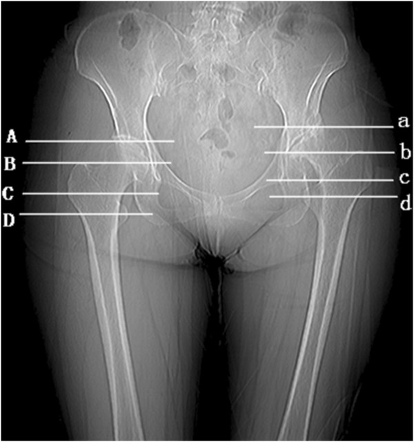
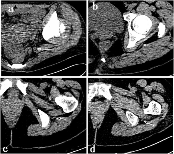
A total of 35 adult patients with unilateral DDH were enrolled in this retrospective study. We reviewed the patients' computed tomography (CT) scans, which included the area from the iliac crest to below the lesser trochanter. The distance between the sciatic nerve and regional anatomic landmarks in four different sections on CT scans was measured to identify the course of the sciatic nerve.
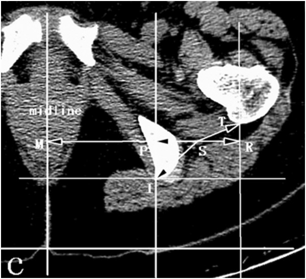
The distance from the sciatic nerve to the spine's midline was shorter on the affected side than on the healthy side (p < 0.05); the same difference was also detected in the distance to the ilium/ischium outside the true pelvis (p < 0.05). The distance to the greater trochanter was longer on the affected side (p < 0.05). However, the two sides showed no significant difference in the distance from the sciatic nerve to the lesser trochanter (p > 0.05).
For patients with unilateral DDH, the sciatic nerve was located near the ischium and ilium but relatively far from the femur of the affected hip joint, compared to its location on the healthy side. These findings reveal that sciatic nerve becomes shorter in the affected low-limb and is relatively unlikely to be directly injuried using the posterolateral approach in patients with unilateral DDH.
坐骨神经损伤是手术中灾难性的不良并发症,可导致使人衰弱的疼痛、功能障碍及生活质量下降。发育性髋关节发育不良(DDH)患者在全髋关节置换术(THA)后发生坐骨神经损伤的发生率较高。更好地了解DDH患者坐骨神经的走行可能有助于降低THA后坐骨神经损伤的风险。
本回顾性研究共纳入35例单侧DDH成年患者。我们回顾了患者的计算机断层扫描(CT)图像,其范围包括从髂嵴至小转子下方区域。测量CT图像上四个不同层面坐骨神经与局部解剖标志之间的距离,以确定坐骨神经的走行。
患侧坐骨神经至脊柱中线的距离短于健侧(p<0.05);至真骨盆外髂骨/坐骨的距离也存在同样差异(p<0.05)。患侧坐骨神经至大转子的距离较长(p<0.05)。然而,两侧坐骨神经至小转子的距离无显著差异(p>0.05)。
对于单侧DDH患者,与健侧相比,患侧坐骨神经位于坐骨和髂骨附近,但离患侧髋关节的股骨相对较远。这些发现表明,患侧下肢的坐骨神经变短,采用后外侧入路时,单侧DDH患者的坐骨神经相对不太可能直接受到损伤。