Passow Susanne, Specht Karsten, Adamsen Tom Christian, Biermann Martin, Brekke Njål, Craven Alexander Richard, Ersland Lars, Grüner Renate, Kleven-Madsen Nina, Kvernenes Ole-Heine, Schwarzlmüller Thomas, Olesen Rasmus Aamand, Hugdahl Kenneth
Department of Biological and Medical Psychology, University of Bergen, Bergen, Norway; NORMENT Center of Excellence, University of Oslo, Norway.
Hum Brain Mapp. 2015 Jun;36(6):2027-38. doi: 10.1002/hbm.22753. Epub 2015 Feb 3.
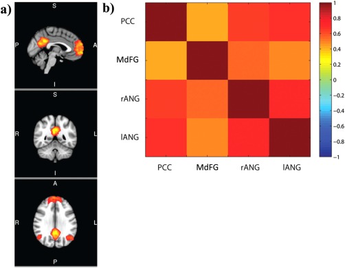
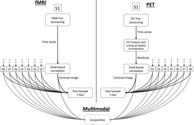
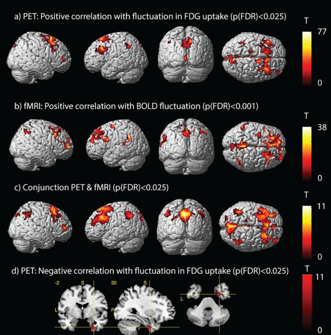
Over the last decade, the brain's default-mode network (DMN) and its function has attracted a lot of attention in the field of neuroscience. However, the exact underlying mechanisms of DMN functional connectivity, or more specifically, the blood-oxygen level-dependent (BOLD) signal, are still incompletely understood. In the present study, we combined 2-deoxy-2-[(18) F]fluoroglucose positron emission tomography (FDG-PET), proton magnetic resonance spectroscopy ((1) H-MRS), and resting-state functional magnetic resonance imaging (rs-fMRI) to investigate more directly the association between local glucose consumption, local glutamatergic neurotransmission and DMN functional connectivity during rest. The results of the correlation analyzes using the dorsal posterior cingulate cortex (dPCC) as seed region showed spatial similarities between fluctuations in FDG-uptake and fluctuations in BOLD signal. More specifically, in both modalities the same DMN areas in the inferior parietal lobe, angular gyrus, precuneus, middle, and medial frontal gyrus were positively correlated with the dPCC. Furthermore, we could demonstrate that local glucose consumption in the medial frontal gyrus, PCC and left angular gyrus was associated with functional connectivity within the DMN. We did not, however, find a relationship between glutamatergic neurotransmission and functional connectivity. In line with very recent findings, our results lend further support for a close association between local metabolic activity and functional connectivity and provide further insights towards a better understanding of the underlying mechanism of the BOLD signal.
在过去十年中,大脑的默认模式网络(DMN)及其功能在神经科学领域引起了广泛关注。然而,DMN功能连接的确切潜在机制,或者更具体地说,血氧水平依赖(BOLD)信号,仍未完全明确。在本研究中,我们结合了2-脱氧-2-[(18)F]氟葡萄糖正电子发射断层扫描(FDG-PET)、质子磁共振波谱((1)H-MRS)和静息态功能磁共振成像(rs-fMRI),以更直接地研究静息状态下局部葡萄糖消耗、局部谷氨酸能神经传递与DMN功能连接之间的关联。以背侧后扣带回皮质(dPCC)作为种子区域进行相关性分析的结果显示,FDG摄取波动与BOLD信号波动之间存在空间相似性。更具体地说,在这两种模式下,顶下小叶、角回、楔前叶、额中回和额内侧回中的相同DMN区域与dPCC呈正相关。此外,我们能够证明额内侧回、PCC和左侧角回中的局部葡萄糖消耗与DMN内的功能连接有关。然而,我们并未发现谷氨酸能神经传递与功能连接之间存在关联。与最近的研究结果一致,我们的结果进一步支持了局部代谢活动与功能连接之间的密切关联,并为更好地理解BOLD信号的潜在机制提供了进一步的见解。