Nishida Kiyotaka, Ubukata Hideyuki, Konishi Satoru, Shimazaki Jiro, Yano Youko, Morishita Yukio, Tabuchi Takafumi
Department of Gastroenterological Surgery, Tokyo Medical University, Ibaraki Medical Center, 3-20-1 Chuo Ami, Inashiki, Ibaraki, 300-0395, Japan.
Department of Diagnostic Pathology Division, Tokyo Medical University, Ibaraki Medical Center, 3-20-1 Chuo Ami, Inashiki, Ibaraki, 300-0395, Japan.
World J Surg Oncol. 2015 Feb 4;13:17. doi: 10.1186/s12957-014-0422-4.
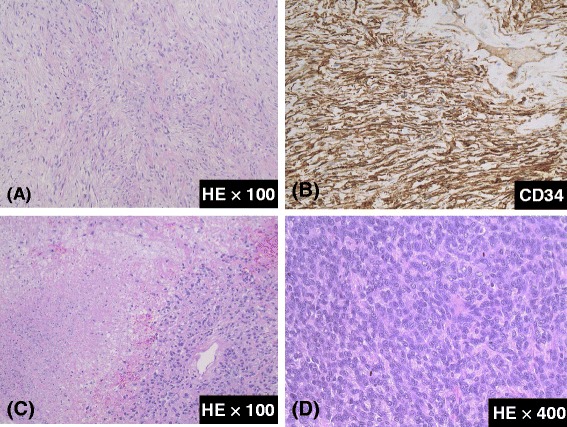
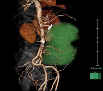
We report on an extremely rare case of a giant solitary fibrous tumor (SFT) of the mesentery in a 65-year-old male who was admitted to our hospital because of lower abdominal pain and abdominal fullness. Computed tomography demonstrated a well-defined solid mass of 25 × 11 cm located in the lower abdomen, which was completely resected during surgery. Histopathologically, this lesion had a heterogeneous cell population, mainly comprising spindle cells with fibrous collagen proliferation, and various other cell populations exhibiting patternless growth. Immunohistochemically, the tumor revealed strong and diffuse staining for CD34, bcl-2, and vimentin, and a high mitotic index (seven mitoses per 10 high-power fields). We diagnosed this case as an SFT of the mesentery, which is unusual according to a PubMed search that reported only nine such cases. Our case may be the largest tumor reported to date, and only one retrieved case reported recurrence, although the lesion was exceptionally large with deep invasion. Nonetheless, the lesion in our case was larger than that in the reported case of recurrence and invasive to the ileum. Since surgery, there has been no evidence of recurrence. Hence, we propose that a large SFT and high mitotic index may present risk factors for recurrence. Therefore, long-term careful follow-up is necessary in such cases, although our case exhibited few risk factors for recurrence. A follow-up at 12 months after surgery found no indications of recurrence.
我们报告了一例极为罕见的肠系膜巨大孤立性纤维瘤(SFT)病例,患者为一名65岁男性,因下腹部疼痛和腹胀入院。计算机断层扫描显示下腹部有一个边界清晰的实性肿块,大小为25×11厘米,手术中完整切除。组织病理学检查显示,该病变细胞群体异质性,主要由伴有纤维胶原增生的梭形细胞组成,还有其他各种细胞群体呈无规律生长。免疫组织化学检查显示,肿瘤对CD34、bcl-2和波形蛋白呈强阳性弥漫性染色,有较高的有丝分裂指数(每10个高倍视野中有7个有丝分裂)。我们将此病例诊断为肠系膜SFT,根据PubMed搜索,仅报道了9例此类病例,本病例并不常见。我们的病例可能是迄今为止报道的最大肿瘤,尽管病变异常大且侵犯较深,但检索到的病例中只有1例报道有复发。尽管如此,我们病例中的病变比报道的复发病例更大,且侵犯到了回肠。自手术以来,没有复发的迹象。因此,我们认为大的SFT和高有丝分裂指数可能是复发的危险因素。因此,尽管我们的病例复发危险因素较少,但此类病例仍有必要进行长期密切随访。术后12个月的随访未发现复发迹象。