Huma Zilli, Maxwell David J
Spinal Cord Group, Institute of Neuroscience and Psychology, College of Medicine, Veterinary Medicine and Life Sciences, University of Glasgow Glasgow, UK.
Front Neuroanat. 2015 Jan 22;9:1. doi: 10.3389/fnana.2015.00001. eCollection 2015.
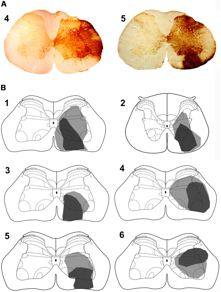
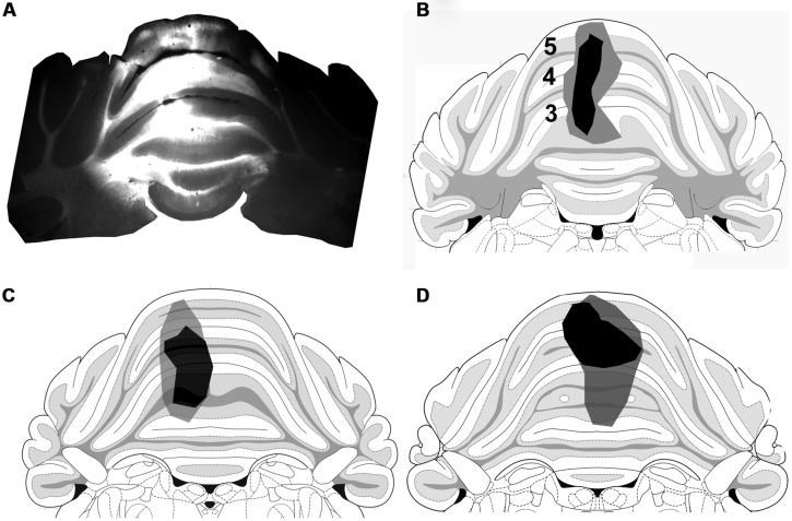
In addition to classical spinocerebellar pathways, the cerebellum receives information from the spinal cord indirectly via spino-bulbar-cerebellar systems. One of the structures in this pathway is the lateral reticular nucleus (LRt). We performed series of experiments to investigate the organization and neurotransmitter content of spinoreticular tract (SRT) neurons in the lumbar spinal cord that project to the LRt. Three rats received injections of the b subunit of Cholera toxin (CTb) or Fluorogold (FG) within the left and right LRt. The majority of SRT cells (56-61%) were found within the contralateral medial intermediate gray matter where small numbers (7-10%) of double-labeled cells were also present on both sides of the cord. Six rats received unilateral spinal injections of CTb to label spinal projections to the LRt. Injections of FG were made also into the anterior lobe of the cerebellum to label LRt pre-cerebellar neurons. Terminals were found mainly ipsilateral to spinal injection sites within the central and ventrolateral regions of the LRt. Immunocytochemical analysis of SRT terminals revealed that the majority (75%) were contained vesicular glutamate transporter 2 but a minority (20%) contained the vesicular GABA transporter. The inhibitory subpopulation was found to be GABAergic, glycinergic, or contained both transmitters. Inhibitory and excitatory terminals were present within overlapping regions of the nucleus. Most CTb terminals contacting LRt pre-cerebellar neurons were excitatory (80%) whereas a minority were inhibitory and most cells (88%) received contacts from both inhibitory and excitatory terminals. This study shows that SRT axons in the LRt have the capacity to exert direct excitatory and inhibitory actions on LRt pre-cerebellar neurons. Thus spinal cord input has the capacity to facilitate or depress the activity of individual LRt cells which in turn adjust activity in the cerebellum to produce coordinated motor behaviors.
除了经典的脊髓小脑通路外,小脑还通过脊髓 - 延髓 - 小脑系统间接从脊髓接收信息。该通路中的结构之一是外侧网状核(LRt)。我们进行了一系列实验,以研究投射至LRt的腰段脊髓脊髓网状束(SRT)神经元的组织和神经递质含量。三只大鼠在左右LRt内注射霍乱毒素(CTb)的b亚基或荧光金(FG)。大多数SRT细胞(56 - 61%)位于对侧内侧中间灰质内,脊髓两侧也有少量(7 - 10%)双标记细胞。六只大鼠接受单侧脊髓注射CTb以标记投射至LRt的脊髓投射纤维。同时也向小脑前叶注射FG以标记LRt小脑前神经元。终末主要位于脊髓注射部位同侧的LRt中央和腹外侧区域。对SRT终末的免疫细胞化学分析显示,大多数(75%)含有囊泡谷氨酸转运体2,但少数(20%)含有囊泡GABA转运体。抑制性亚群被发现为GABA能、甘氨酸能或同时含有这两种递质。抑制性和兴奋性终末存在于核的重叠区域内。大多数与LRt小脑前神经元接触的CTb终末是兴奋性的(80%),而少数是抑制性的,并且大多数细胞(88%)接受来自抑制性和兴奋性终末的接触。这项研究表明,LRt中的SRT轴突有能力对LRt小脑前神经元施加直接的兴奋性和抑制性作用。因此,脊髓输入有能力促进或抑制单个LRt细胞的活动,进而调节小脑的活动以产生协调的运动行为。