Talaie Tara, Pratt Stephen J P, Vanegas Camilo, Xu Su, Henn R Frank, Yarowsky Paul, Lovering Richard M
Department of Orthopaedics, University of Maryland School of Medicine.
Department of Diagnostic Radiology and Nuclear Medicine, University of Maryland School of Medicine.
Orthop J Sports Med. 2015 Jan 22;3(1). doi: 10.1177/2325967114566185.
Muscle strains are one of the most common injuries treated by physicians. Standard conservative therapy for acute muscle strains usually involves short-term rest, ice, and non-steroidal anti-inflammatory medications, but there is no clear consensus regarding treatments to accelerate recovery. Recently, clinical use of platelet-rich plasma (PRP) has gained momentum as an option for therapy and is appealing for many reasons, most notably because it provides growth factors in physiological proportions and it is autologous, safe, easily accessible, and potentially beneficial. Local delivery of patients' PRP to injured muscles can hasten recovery of function. However, specific targeting of PRP to sites of tissue damage in vivo is a major challenge that can limit its efficacy.
Location of PRP delivery can be monitored and controlled in vivo with non-invasive tools.
Controlled laboratory study using rats.
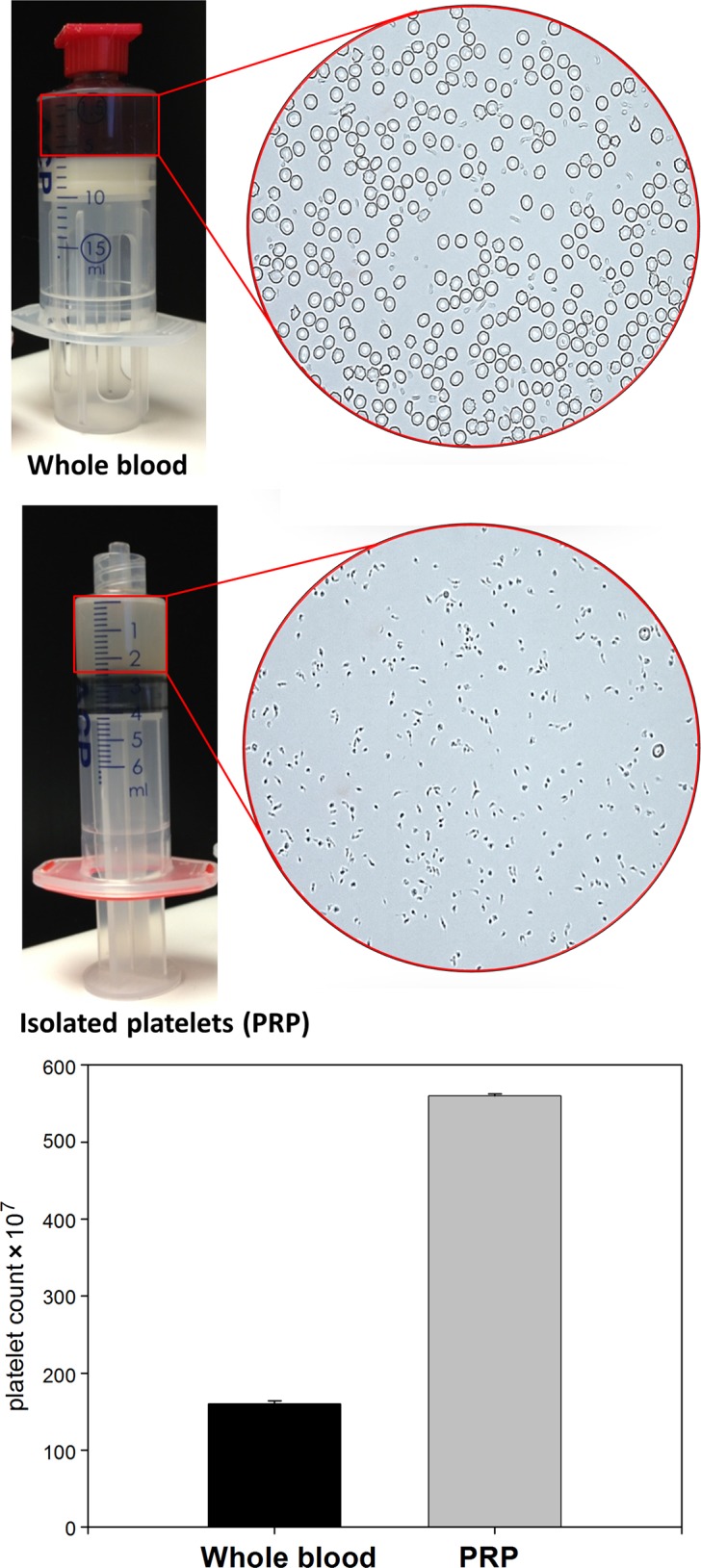
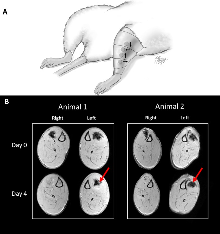
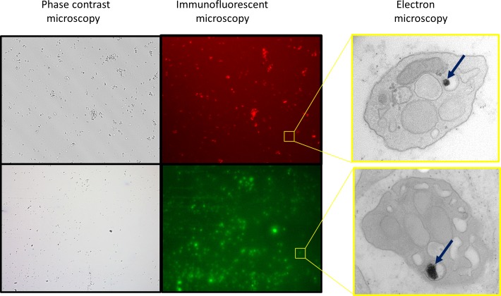
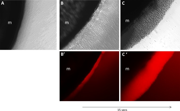
Superparamagnetic iron oxide nanoparticles (SPIONs) can be visualized by both MRI (in vivo) and fluorescence microscopy (after tissue harvesting). We labeled PRP with SPIONs and administered intramuscular injections of SPION-containing platelets. MRI was used to monitor the ability to manipulate and retain the location of PRP in vivo by placement of an external magnet. Platelets were isolated from whole blood and incubated with SPIONs. Following SPION incubation with PRP, a magnetic field was used to manipulate platelet location in culture dishes. In vivo, the tibialis anterior muscles (TAs) of anesthetized Sprague-Dawley rats were injected with SPION-containing platelets and MRI was used to track platelet position with and without a magnet worn over the TAs for 4 days.
The method used to isolate PRP yielded a high concentration (almost 4-fold increase) of platelets. In vitro experiments show that the platelets successfully took up SPIONs and then rapidly responded to an applied magnetic field. Platelets without SPIONs did not respond to the magnetic field. In vivo experiments show that the SPION-containing platelets can be non-invasively maintained at a specific site with the application of a magnetic field.
PRP may be a useful product in clinical treatment of muscle injuries, but one problem with using PRP as a therapeutic tool, is retaining PRP at the site of injury. We propose a potential solution with our findings that support this method at the cell, whole muscle, and in vivo levels. Controlling the location of PRP will allow the clustering of PRP to enrich the target area with growth factors and will prevent loss of the platelets over time at the site of injury.
肌肉拉伤是医生治疗的最常见损伤之一。急性肌肉拉伤的标准保守治疗通常包括短期休息、冰敷和使用非甾体类抗炎药物,但对于加速恢复的治疗方法尚无明确共识。最近,富血小板血浆(PRP)作为一种治疗选择在临床上的应用越来越广泛,其具有吸引力的原因有很多,最显著的是它能以生理比例提供生长因子,并且是自体的、安全的、易于获取且可能有益的。将患者的PRP局部注射到受伤肌肉中可以加速功能恢复。然而,在体内将PRP特异性靶向组织损伤部位是一个重大挑战,这可能会限制其疗效。
可以使用非侵入性工具在体内监测和控制PRP的递送位置。
使用大鼠进行的对照实验室研究。
超顺磁性氧化铁纳米颗粒(SPIONs)可通过MRI(体内)和荧光显微镜(组织采集后)进行可视化。我们用SPIONs标记PRP,并进行含SPIONs血小板的肌肉注射。通过放置外部磁体,利用MRI监测在体内操纵和保持PRP位置的能力。从全血中分离血小板,并与SPIONs孵育。在PRP与SPIONs孵育后,使用磁场在培养皿中操纵血小板位置。在体内,对麻醉的Sprague-Dawley大鼠的胫前肌(TA)注射含SPIONs的血小板,并使用MRI在TA上佩戴或不佩戴磁体的情况下追踪血小板位置,持续4天。
用于分离PRP的方法产生了高浓度(几乎增加4倍)的血小板。体外实验表明,血小板成功摄取了SPIONs,然后对施加的磁场迅速做出反应。不含SPIONs的血小板对磁场无反应。体内实验表明,通过施加磁场,含SPIONs的血小板可以非侵入性地维持在特定部位。
PRP可能是肌肉损伤临床治疗中的一种有用产品,但将PRP用作治疗工具的一个问题是如何将其保留在损伤部位。我们通过在细胞、全肌肉和体内水平上支持该方法的研究结果提出了一种潜在的解决方案。控制PRP的位置将使PRP聚集,从而用生长因子富集靶区域,并防止血小板在损伤部位随时间流失。