• 文献检索
  • 文档翻译
  • 深度研究
  • 学术资讯
  • Suppr Zotero 插件Zotero 插件
  • 邀请有礼
  • 套餐&价格
  • 历史记录
应用&插件
Suppr Zotero 插件Zotero 插件浏览器插件Mac 客户端Windows 客户端微信小程序
定价
高级版会员购买积分包购买API积分包
服务
文献检索文档翻译深度研究API 文档MCP 服务
关于我们
关于 Suppr公司介绍联系我们用户协议隐私条款
关注我们

Suppr 超能文献

核心技术专利:CN118964589B侵权必究
粤ICP备2023148730 号-1Suppr @ 2026

文献检索

告别复杂PubMed语法,用中文像聊天一样搜索,搜遍4000万医学文献。AI智能推荐,让科研检索更轻松。

立即免费搜索

文件翻译

保留排版,准确专业,支持PDF/Word/PPT等文件格式,支持 12+语言互译。

免费翻译文档

深度研究

AI帮你快速写综述,25分钟生成高质量综述,智能提取关键信息,辅助科研写作。

立即免费体验

术中三维成像(O型臂)用于评估椎弓根螺钉位置:它能否防止不可接受的螺钉置入?

Intraoperative 3-dimensional imaging (O-arm) for assessment of pedicle screw position: Does it prevent unacceptable screw placement?

作者信息

Sembrano Jonathan N, Polly David W, Ledonio Charles Gerald T, Santos Edward Rainier G

机构信息

Department of Orthopaedic Surgery, University of Minnesota, Minneapolis, MN ; Minneapolis VA Health Care System, Minneapolis, MN.

Department of Orthopaedic Surgery, University of Minnesota, Minneapolis, MN.

出版信息

Int J Spine Surg. 2012 Dec 1;6:49-54. doi: 10.1016/j.ijsp.2011.11.002. eCollection 2012.

DOI:10.1016/j.ijsp.2011.11.002
PMID:25694871
原文链接:https://pmc.ncbi.nlm.nih.gov/articles/PMC4300877/
Abstract

BACKGROUND

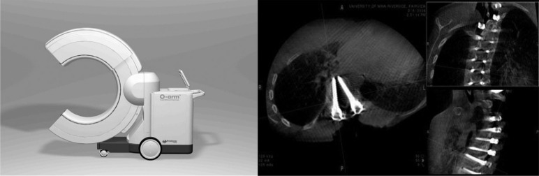
Pedicle screws are biomechanically superior over other spinal fixation devices. When improperly positioned, they lose this advantage and put adjacent structures at risk. Accurate placement is therefore critical. Postoperative computed tomography (CT) scans are the imaging gold standard and have shown malposition rates ranging from 2% to 41%. The O-arm (Medtronic Navigation, Louisville, Colorado) is an intraoperative CT scanner that may allow intervention for malpositioned screws while patients are still in the operating room. However, this has not yet been shown in clinical studies. The primary objective of this study was to assess the usefulness of the O-arm for evaluating pedicle screw position by answering the following question: What is the rate of intraoperative pedicle screw revision brought about by O-arm imaging information? A secondary question was also addressed: What is the rate of unacceptable thoracic and lumbar pedicle screw placement as assessed by intraoperative O-arm imaging?

METHODS

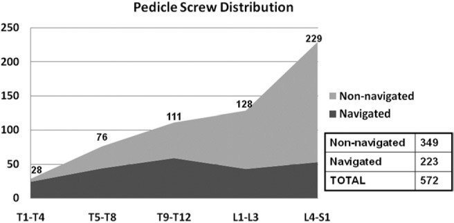
This is a case series of consecutive patients who have undergone spine surgery for which an intraoperative 3-dimensional (3D) CT scan was used to assess pedicle screw position. The study comprised 602 pedicle screws (235 thoracic and 367 lumbar/sacral) placed in 76 patients, and intraoperative 3D (O-arm) imaging was obtained to assess screw position. Action taken at the time of surgery based on imaging information was noted. An independent review of all scans was also conducted, and all screws were graded as either optimal (no breach), acceptable (breach ≤2 mm), or unacceptable (breach >2 mm). The rate of pedicle screw revision, as detected by intraoperative 3D CT scan, was determined.

RESULTS

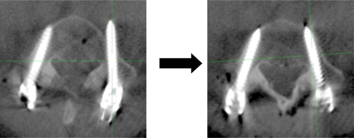
On the basis of 3D imaging information, 17 of 602 screws (2.8%) in 14 of 76 cases (18.4%) were revised at the time of surgery. On independent review of multiplanar images, 11 screws (1.8%) were found to be unacceptable, 32 (5.3%) were acceptable, and 559 (92.9%) were optimal. All unacceptable screws were revised to an optimal or acceptable position, and an additional 6 acceptable screws were revised to an optimal position. Thus, by the end of the cases, none of the 602 pedicle screws in the 76 surgical procedures was in an unacceptable position.

CONCLUSION

The new-generation intraoperative 3D imaging system (O-arm) is a useful tool that allows more accurate assessment of pedicle screw position than plain radiographs or fluoroscopy alone. It prompted intraoperative repositioning of 2.8% of pedicle screws in our series. Most importantly, it allowed identification and revision of all unacceptably placed pedicle screws without the need for reoperation.

摘要

背景

椎弓根螺钉在生物力学方面优于其他脊柱固定装置。若位置不当,它们将失去这一优势,并使相邻结构处于危险之中。因此,准确放置至关重要。术后计算机断层扫描(CT)是成像的金标准,其显示的置钉位置不当率在2%至41%之间。O型臂(美敦力导航公司,科罗拉多州路易斯维尔)是一种术中CT扫描仪,它可能允许在患者仍在手术室时对位置不当的螺钉进行干预。然而,临床研究尚未证实这一点。本研究的主要目的是通过回答以下问题来评估O型臂在评估椎弓根螺钉位置方面的实用性:O型臂成像信息导致的术中椎弓根螺钉翻修率是多少?还提出了一个次要问题:术中O型臂成像评估的胸腰椎椎弓根螺钉放置不可接受率是多少?

方法

这是一系列连续接受脊柱手术的患者病例,术中使用三维(3D)CT扫描评估椎弓根螺钉位置。该研究包括76例患者置入的602枚椎弓根螺钉(235枚胸椎和367枚腰椎/骶椎),并获得术中3D(O型臂)成像以评估螺钉位置。记录基于成像信息在手术时采取的行动。还对所有扫描进行了独立审查,所有螺钉被分为最佳(无突破)、可接受(突破≤2 mm)或不可接受(突破>2 mm)。确定术中3D CT扫描检测到的椎弓根螺钉翻修率。

结果

根据3D成像信息,76例中的14例(18.4%)的602枚螺钉中有17枚(2.8%)在手术时进行了翻修。在对多平面图像进行独立审查时,发现11枚螺钉(1.8%)不可接受,32枚(5.3%)可接受,559枚(92.9%)最佳。所有不可接受的螺钉均被翻修至最佳或可接受位置,另外6枚可接受的螺钉被翻修至最佳位置。因此,在这些病例结束时,76例手术中的602枚椎弓根螺钉均未处于不可接受的位置。

结论

新一代术中3D成像系统(O型臂)是一种有用的工具,与单纯的平片或透视相比,它能更准确地评估椎弓根螺钉位置。在我们的系列研究中,它促使2.8%的椎弓根螺钉在术中重新定位。最重要的是,它能够识别并翻修所有放置位置不可接受的椎弓根螺钉,而无需再次手术。

https://cdn.ncbi.nlm.nih.gov/pmc/blobs/2fe7/4300877/bcf7ac51fcdc/IJSS-6-2011-11-002-g003.jpg
https://cdn.ncbi.nlm.nih.gov/pmc/blobs/2fe7/4300877/4660473ba7d9/IJSS-6-2011-11-002-g001.jpg
https://cdn.ncbi.nlm.nih.gov/pmc/blobs/2fe7/4300877/f6da05238e2b/IJSS-6-2011-11-002-g002.jpg
https://cdn.ncbi.nlm.nih.gov/pmc/blobs/2fe7/4300877/bcf7ac51fcdc/IJSS-6-2011-11-002-g003.jpg
https://cdn.ncbi.nlm.nih.gov/pmc/blobs/2fe7/4300877/4660473ba7d9/IJSS-6-2011-11-002-g001.jpg
https://cdn.ncbi.nlm.nih.gov/pmc/blobs/2fe7/4300877/f6da05238e2b/IJSS-6-2011-11-002-g002.jpg
https://cdn.ncbi.nlm.nih.gov/pmc/blobs/2fe7/4300877/bcf7ac51fcdc/IJSS-6-2011-11-002-g003.jpg

相似文献

1
Intraoperative 3-dimensional imaging (O-arm) for assessment of pedicle screw position: Does it prevent unacceptable screw placement?术中三维成像(O型臂)用于评估椎弓根螺钉位置:它能否防止不可接受的螺钉置入?
Int J Spine Surg. 2012 Dec 1;6:49-54. doi: 10.1016/j.ijsp.2011.11.002. eCollection 2012.
2
Use of the Airo mobile intraoperative CT system versus the O-arm for transpedicular screw fixation in the thoracic and lumbar spine: a retrospective cohort study of 263 patients.在胸椎和腰椎经椎弓根螺钉固定中使用Airo移动术中CT系统与O型臂的比较:一项对263例患者的回顾性队列研究。
J Neurosurg Spine. 2018 Oct;29(4):397-406. doi: 10.3171/2018.1.SPINE17927. Epub 2018 Jul 6.
3
A prospective multicenter registry on the accuracy of pedicle screw placement in the thoracic, lumbar, and sacral levels with the use of the O-arm imaging system and StealthStation Navigation.后路椎弓根螺钉置入准确性的前瞻性多中心研究:O 臂影像系统与 StealthStation 导航系统的应用
Spine (Phila Pa 1976). 2012 Dec 1;37(25):E1580-7. doi: 10.1097/BRS.0b013e318271b1fa.
4
Pediatric pedicle screw placement using intraoperative computed tomography and 3-dimensional image-guided navigation.术中计算机断层扫描和三维图像引导导航在儿科椎弓根螺钉植入中的应用。
Spine (Phila Pa 1976). 2012 Feb 1;37(3):E188-94. doi: 10.1097/BRS.0b013e31822a2e0a.
5
Fluoroscopy-Assisted Freehand Versus 3D-Navigated Imaging-Assisted Pedicle Screw Insertion: A Multicenter Study.透视引导徒手与 3D 导航影像辅助椎弓根螺钉置入的比较:一项多中心研究。
Acta Neurochir Suppl. 2023;135:425-430. doi: 10.1007/978-3-031-36084-8_65.
6
Electromyography stimulation compared with intraoperative O-arm imaging for evaluating pedicle screw breaches in lumbar spine surgery: a prospective analysis of 1006 screws in 164 patients.肌电图刺激与术中 O 臂成像在腰椎手术中评估椎弓根螺钉穿透的比较:164 例患者 1006 枚螺钉的前瞻性分析。
Spine J. 2019 Feb;19(2):206-211. doi: 10.1016/j.spinee.2018.06.353. Epub 2018 Jun 28.
7
Safety and Accuracy of Freehand Pedicle Screw Placement and the Role of Intraoperative O-Arm: A Single-Institution Experience.徒手椎弓根螺钉置入的安全性和准确性以及术中O型臂的作用:单机构经验
Spine (Phila Pa 1976). 2023 Feb 1;48(3):180-188. doi: 10.1097/BRS.0000000000004497. Epub 2022 Sep 28.
8
Benefit and accuracy of intraoperative 3D-imaging after pedicle screw placement: a prospective study in stabilizing thoracolumbar fractures.椎弓根螺钉置入术后术中三维成像的益处与准确性:一项关于胸腰椎骨折固定的前瞻性研究
Eur Spine J. 2009 Oct;18(10):1469-77. doi: 10.1007/s00586-009-1050-5. Epub 2009 Jun 10.
9
Intraoperative Computed Tomography-Guided Navigation for Pediatric Spine Patients Reduced Return to Operating Room for Screw Malposition Compared With Freehand/Fluoroscopic Techniques.与徒手/透视技术相比,术中计算机断层扫描引导导航用于小儿脊柱患者可减少因螺钉位置不当而返回手术室的情况。
Spine Deform. 2019 Jul;7(4):577-581. doi: 10.1016/j.jspd.2018.11.012.
10
The accuracy of intraoperative O-arm images for the assessment of pedicle screw postion.术中 O 臂图像评估椎弓根螺钉位置的准确性。
Spine (Phila Pa 1976). 2012 Jan 15;37(2):E119-25. doi: 10.1097/BRS.0b013e3182257cae.

引用本文的文献

1
Feature-based multimodal 3D/2D and 3D/3D registration framework for pedicle screw registration and evaluation.基于特征的多模态3D/2D和3D/3D配准框架用于椎弓根螺钉配准与评估。
Eur Spine J. 2025 Sep 13. doi: 10.1007/s00586-025-09342-6.
2
Comparison of accuracy of pedicle screw placement for adolescent idiopathic scoliosis using freehand fluoroscopic, navigation, and robotic-assisted techniques - a systematic review and bayesian network meta-analysis.使用徒手透视、导航和机器人辅助技术进行青少年特发性脊柱侧弯椎弓根螺钉置入准确性的比较——一项系统评价和贝叶斯网络荟萃分析
Neurosurg Rev. 2025 Feb 21;48(1):257. doi: 10.1007/s10143-025-03333-3.
3

本文引用的文献

1
Clinical assessment of percutaneous lumbar pedicle screw placement using theO-arm multidimensional surgical imaging system.经皮腰椎椎弓根螺钉置入的临床评估:O 臂多维手术成像系统的应用。
Neurosurgery. 2012 Apr;70(4):990-5. doi: 10.1227/NEU.0b013e318237a829.
2
Pediatric pedicle screw placement using intraoperative computed tomography and 3-dimensional image-guided navigation.术中计算机断层扫描和三维图像引导导航在儿科椎弓根螺钉植入中的应用。
Spine (Phila Pa 1976). 2012 Feb 1;37(3):E188-94. doi: 10.1097/BRS.0b013e31822a2e0a.
3
The accuracy of intraoperative O-arm images for the assessment of pedicle screw postion.
O-arm navigation without apnoea in thoracolumbar and lumbar spine surgery: Outcomes and considerations in a prospective study.
胸腰椎和腰椎手术中无需呼吸暂停的O型臂导航:一项前瞻性研究的结果与考量
J Clin Orthop Trauma. 2024 Sep 30;57:102551. doi: 10.1016/j.jcot.2024.102551. eCollection 2024 Oct.
4
Robot-assisted pedicle screw insertion versus navigation-based and freehand techniques for posterior spinal fusion in scoliosis: a systematic review and meta-analysis.机器人辅助椎弓根螺钉置入与导航辅助和徒手技术在后路脊柱融合治疗脊柱侧凸中的比较:系统评价和荟萃分析。
Spine Deform. 2024 Sep;12(5):1203-1215. doi: 10.1007/s43390-024-00879-y. Epub 2024 Apr 15.
5
Intraoperative 3D Imaging Reduces Pedicle Screw Related Complications and Reoperations in Adolescents Undergoing Posterior Spinal Fusion for Idiopathic Scoliosis: A Retrospective Study.术中三维成像减少特发性脊柱侧凸后路脊柱融合术青少年患者椎弓根螺钉相关并发症及再次手术:一项回顾性研究
Children (Basel). 2022 Jul 28;9(8):1129. doi: 10.3390/children9081129.
6
A Hybrid 3D-2D Image Registration Framework for Pedicle Screw Trajectory Registration between Intraoperative X-ray Image and Preoperative CT Image.一种用于术中X射线图像与术前CT图像之间椎弓根螺钉轨迹配准的混合3D-2D图像配准框架。
J Imaging. 2022 Jul 6;8(7):185. doi: 10.3390/jimaging8070185.
7
The Transverse Process Trajectory Technique: An Alternative for Thoracic Pedicle Screw Implantation-Radiographic and Biomechanical Analysis.横突轨迹技术:一种胸椎椎弓根螺钉植入的替代方法——影像学及生物力学分析
Int J Spine Surg. 2021 Apr;15(2):315-323. doi: 10.14444/8041. Epub 2021 Apr 1.
8
Narrative review of intraoperative image guidance for transforaminal lumbar interbody fusion.经椎间孔腰椎椎间融合术中术中影像引导的叙述性综述。
Ann Transl Med. 2021 Jan;9(1):89. doi: 10.21037/atm-20-1971.
9
Transpedicular Screw Placement Accuracy Using the O-Arm Versus Freehand Technique at a Single Institution.在单一机构中使用O型臂与徒手技术进行椎弓根螺钉置入的准确性
Global Spine J. 2022 Apr;12(3):447-451. doi: 10.1177/2192568220956979. Epub 2020 Oct 1.
10
A learning-based method for online adjustment of C-arm Cone-beam CT source trajectories for artifact avoidance.基于学习的方法,用于在线调整 C 臂锥形束 CT 源轨迹以避免伪影。
Int J Comput Assist Radiol Surg. 2020 Nov;15(11):1787-1796. doi: 10.1007/s11548-020-02249-1. Epub 2020 Aug 25.
术中 O 臂图像评估椎弓根螺钉位置的准确性。
Spine (Phila Pa 1976). 2012 Jan 15;37(2):E119-25. doi: 10.1097/BRS.0b013e3182257cae.
4
Computer tomography assessment of pedicle screw placement in lumbar and sacral spine: comparison between free-hand and O-arm based navigation techniques.计算机断层扫描评估腰椎和骶骨脊柱椎弓根螺钉放置:徒手和 O 臂导航技术的比较。
Eur Spine J. 2011 Jun;20(6):875-81. doi: 10.1007/s00586-010-1683-4. Epub 2011 Jan 21.
5
Delayed perforation of the aorta by a thoracic pedicle screw.一枚胸椎椎弓根螺钉导致的主动脉延迟穿孔
Eur Spine J. 2008 Sep;17 Suppl 2(Suppl 2):S351-4. doi: 10.1007/s00586-008-0715-9. Epub 2008 Jul 10.
6
Delayed presentation of aortic injury by pedicle screws: report of two cases and review of the literature.椎弓根螺钉导致的主动脉损伤延迟表现:两例报告及文献复习
J Vasc Surg. 2008 May;47(5):1074-82. doi: 10.1016/j.jvs.2007.11.005. Epub 2008 Feb 1.
7
Surgical revision rates of hooks versus hybrid versus screws versus combined anteroposterior spinal fusion for adolescent idiopathic scoliosis.青少年特发性脊柱侧凸采用钩、混合、螺钉及前后路联合脊柱融合术的手术翻修率。
Spine (Phila Pa 1976). 2007 Sep 15;32(20):2258-64. doi: 10.1097/BRS.0b013e31814b1ba6.
8
Image guidance in spine surgery.脊柱手术中的影像引导
Orthop Clin North Am. 2007 Jul;38(3):451-61; abstract viii. doi: 10.1016/j.ocl.2007.04.001.
9
Complications of thoracic pedicle screws in scoliosis treatment.脊柱侧弯治疗中胸椎椎弓根螺钉的并发症
Spine (Phila Pa 1976). 2007 Jul 1;32(15):1655-61. doi: 10.1097/BRS.0b013e318074d604.
10
Pedicle screw placement accuracy: a meta-analysis.椎弓根螺钉置入准确性:一项荟萃分析。
Spine (Phila Pa 1976). 2007 Feb 1;32(3):E111-20. doi: 10.1097/01.brs.0000254048.79024.8b.