Singh Shishir, Pawar Mansing
Department of Conservative Dentistry and Endodontics, Government Dental College and Hospital, St. George Hospital compound, D Mello Road, Near GPO, Fort. Mumbai, Maharashtra, India.
Eur J Dent. 2015 Jan-Mar;9(1):133-144. doi: 10.4103/1305-7456.149662.
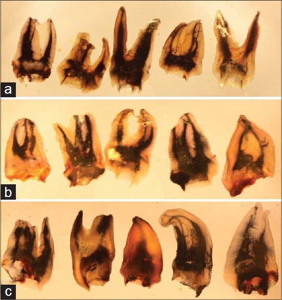
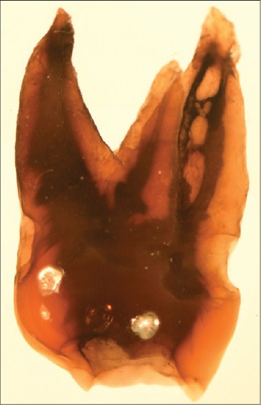
The objective was to study the root canal morphology of South Asian Indian Maxillary molars using a tooth clearing technique.
Hundred teeth each comprising of first, second, and third molars collected from different dental schools and clinics in India were subjected to standard dye penetration, decalcification and clearing procedure before being studied.
The first molar mesiobuccal roots exhibited 69% Type I, 24% Type II, 4% Type IV, 2% Type V, and 1% exhibited a Vertuccis Type VIII canal anatomy. In the group with three separate roots the second molar mesiobuccal roots in exhibited 80.6% Type I, 15.3% Type II, 2.7% Type IV, and 1.4% Type V canal anatomy while the third molars mesiobuccal roots exhibited 57.4% Type I, 32% Type II, 2.1% Type III, 8.5% Type IV, 1% had a Type V canal anatomy in the similar group.
A varied root canal anatomy was seen in the mesiobuccal root canal of the maxillary molars.
采用牙齿透明技术研究南亚印度人上颌磨牙的根管形态。
从印度不同牙科学院和诊所收集的100颗牙齿,每颗牙齿均包含第一、第二和第三磨牙,在进行研究前先进行标准的染料渗透、脱钙和透明处理。
第一磨牙近中颊根呈现69%为I型、24%为II型、4%为IV型、2%为V型,1%呈现Vertucci VIII型根管解剖结构。在有三根独立牙根的组中,第二磨牙近中颊根呈现80.6%为I型、15.3%为II型、2.7%为IV型、1.4%为V型根管解剖结构,而第三磨牙近中颊根在类似组中呈现57.4%为I型、32%为II型、2.1%为III型、8.5%为IV型、1%为V型根管解剖结构。
上颌磨牙近中颊根管呈现出多样的根管解剖结构。