Chung Sook Hyun, Gillies Mark, Sugiyama Yuki, Zhu Ling, Lee So-Ra, Shen Weiyong
Macular Research Group, Clinical Ophthalmology and Eye Health, Save Sight Institute, University of Sydney, Sydney, Australia.
Lens Research Group, Clinical Ophthalmology and Eye Health, Save Sight Institute, University of Sydney, Sydney, Australia.
PLoS One. 2015 Mar 5;10(3):e0118949. doi: 10.1371/journal.pone.0118949. eCollection 2015.
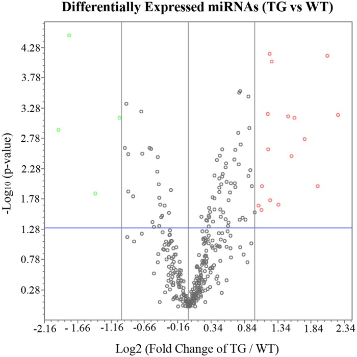
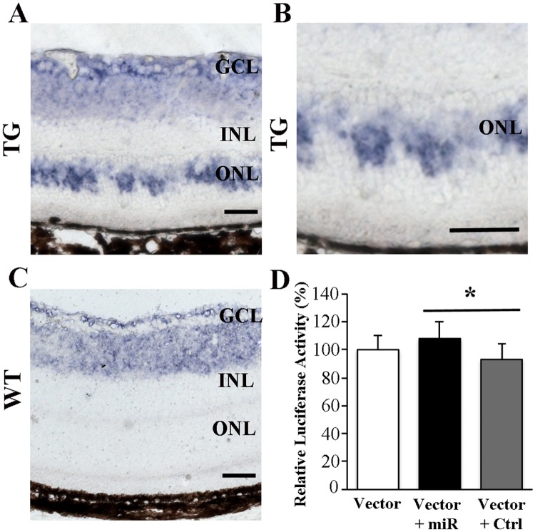
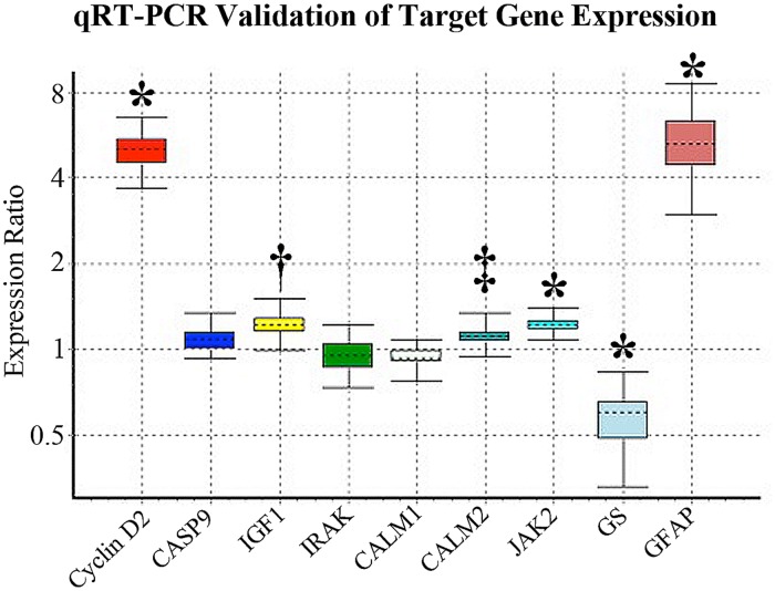
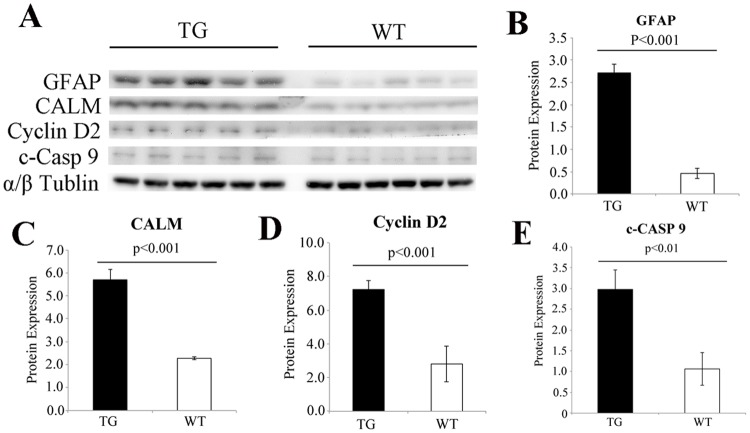
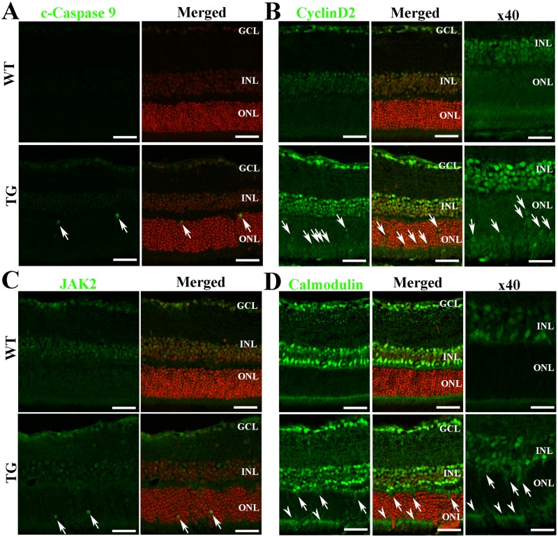
Dysfunction of Müller cells has been implicated in the pathogenesis of several retinal diseases. In order to understand the potential contribution of Müller cells to retinal disease better, we have developed a transgenic model in which foci of Müller cell ablation can be selectively induced. MicroRNAs (miRNAs), small non-coding RNAs that are involved in post-transcriptional modulation, have critical functions in various biological processes. The aim of this study was to profile differential expression of miRNAs and to examine changes in their target genes 2 weeks after Müller cell ablation. We identified 20 miRNAs using the miScript HC PCR array. Data analysis using two target gene prediction databases (TargetScan and mirTarBase) revealed 78 overlapping target genes. DAVID and KEGG pathway analysis suggested that the target genes were generally involved in cell apoptosis, p53, neurotrophin, calcium, chemokine and Jak-STAT signalling pathways. Changes in seven target genes including Cyclin D2, Caspase 9, insulin-like growth factor 1, IL-1 receptor-associated kinase (IRAK), calmodulin (CALM) and Janus kinase 2 (Jak2), were validated with qRT-PCR and western blots. The cellular localisation of cleaved-caspase 9, Cyclin D2, Jak2 and CALM was examined by immunofluorescence studies. We found that the transcription of some miRNAs was positively, rather than negatively, correlated with their target genes. After confirming that overexpressed miR-133a-3p was localised to the outer nuclear layer in the damaged retina, we validated the correlation between miR-133a-3p and one of its predicted target genes, cyclin D2, with a luciferase assay in 661 photoreceptor cells. Results revealed by miRNA profiling, target gene analysis and validation were generally consistent with our previous findings that selective Müller cell ablation causes photoreceptor degeneration and neuroinflammation. Our data on alterations of miRNAs and their target gene expression after Müller cell ablation provide further insights into the potential role of Müller cell dysfunction in retinal disease.
缪勒细胞功能障碍与多种视网膜疾病的发病机制有关。为了更好地理解缪勒细胞对视网膜疾病的潜在影响,我们构建了一种转基因模型,可选择性诱导缪勒细胞消融灶。微小RNA(miRNA)是参与转录后调控的小型非编码RNA,在各种生物学过程中具有关键作用。本研究的目的是分析miRNA的差异表达,并检测缪勒细胞消融2周后其靶基因的变化。我们使用miScript HC PCR阵列鉴定了20种miRNA。使用两个靶基因预测数据库(TargetScan和mirTarBase)进行数据分析,发现了78个重叠的靶基因。DAVID和KEGG通路分析表明,这些靶基因通常参与细胞凋亡、p53、神经营养因子、钙、趋化因子和Jak-STAT信号通路。通过qRT-PCR和蛋白质印迹法验证了包括细胞周期蛋白D2、半胱天冬酶9、胰岛素样生长因子1、白细胞介素-1受体相关激酶(IRAK)、钙调蛋白(CALM)和Janus激酶2(Jak2)在内的7个靶基因的变化。通过免疫荧光研究检测了裂解的半胱天冬酶9、细胞周期蛋白D2、Jak2和CALM的细胞定位。我们发现一些miRNA的转录与其靶基因呈正相关,而非负相关。在确认过表达的miR-133a-3p定位于受损视网膜的外核层后,我们在661个光感受器细胞中通过荧光素酶测定法验证了miR-133a-3p与其预测靶基因之一细胞周期蛋白D2之间的相关性。miRNA谱分析、靶基因分析和验证结果总体上与我们之前的发现一致,即选择性缪勒细胞消融会导致光感受器变性和神经炎症。我们关于缪勒细胞消融后miRNA及其靶基因表达变化的数据,为缪勒细胞功能障碍在视网膜疾病中的潜在作用提供了进一步的见解。