Cramer Julie M, Thompson Timothy, Geskin Albert, LaFramboise William, Lagasse Eric
Department of Pathology, University of Pittsburgh School of Medicine, 200 Lothrop Street, Pittsburgh, PA, 15261, United States of America; McGowan Institute for Regenerative Medicine, University of Pittsburgh, 450 Technology Drive, Suite 300, Pittsburgh, PA, 15219, United States of America.
Department of Bioengineering, University of Pittsburgh, 3700 O'Hara St, Pittsburgh, PA, 15261, United States of America.
PLoS One. 2015 Mar 9;10(3):e0118792. doi: 10.1371/journal.pone.0118792. eCollection 2015.
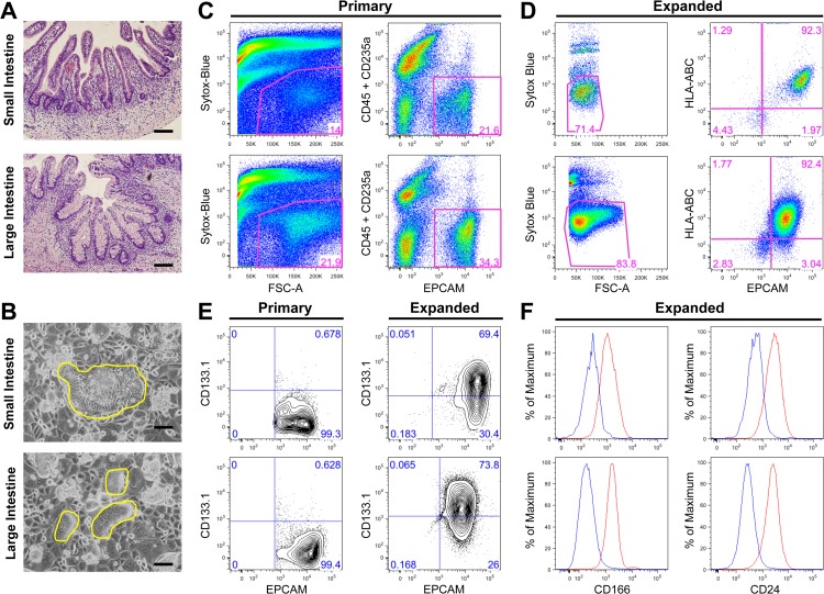
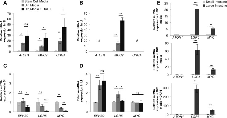
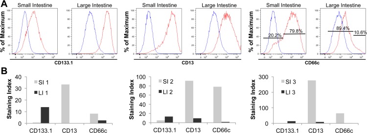
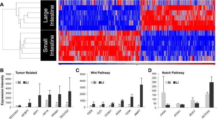
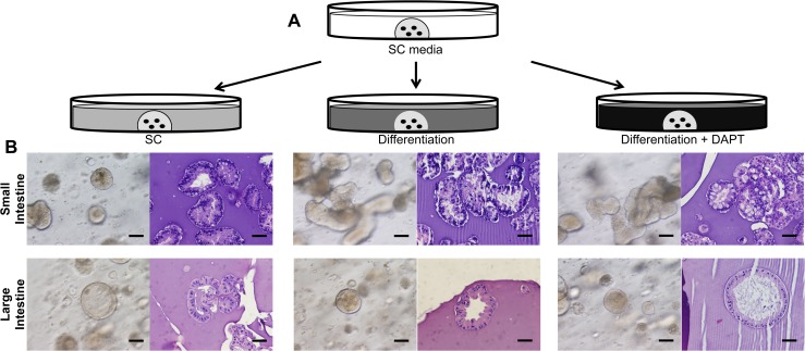
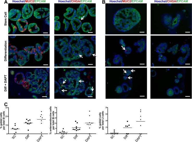
The intestine is composed of an epithelial layer containing rapidly proliferating cells that mature into two regions, the small and the large intestine. Although previous studies have identified stem cells as the cell-of-origin for intestinal epithelial cells, no studies have directly compared stem cells derived from these anatomically distinct regions. Here, we examine intrinsic differences between primary epithelial cells isolated from human fetal small and large intestine, after in vitro expansion, using the Wnt agonist R-spondin 2. We utilized flow cytometry, fluorescence-activated cell sorting, gene expression analysis and a three-dimensional in vitro differentiation assay to characterize their stem cell properties. We identified stem cell markers that separate subpopulations of colony-forming cells in the small and large intestine and revealed important differences in differentiation, proliferation and disease pathways using gene expression analysis. Single cells from small and large intestine cultures formed organoids that reflect the distinct cellular hierarchy found in vivo and respond differently to identical exogenous cues. Our characterization identified numerous differences between small and large intestine epithelial stem cells suggesting possible connections to intestinal disease.
肠道由一层上皮细胞组成,其中包含快速增殖的细胞,这些细胞成熟后形成两个区域,即小肠和大肠。尽管先前的研究已将干细胞确定为肠道上皮细胞的起源细胞,但尚无研究直接比较源自这些解剖学上不同区域的干细胞。在这里,我们使用Wnt激动剂R-spondin 2,研究了体外扩增后人胎儿小肠和大肠分离出的原代上皮细胞之间的内在差异。我们利用流式细胞术、荧光激活细胞分选、基因表达分析和三维体外分化试验来表征它们的干细胞特性。我们鉴定出了可区分小肠和大肠中集落形成细胞亚群的干细胞标志物,并通过基因表达分析揭示了分化、增殖和疾病途径方面的重要差异。来自小肠和大肠培养物的单细胞形成了类器官,这些类器官反映了体内发现的独特细胞层次结构,并且对相同的外源性信号有不同的反应。我们的表征确定了小肠和大肠上皮干细胞之间的许多差异,这表明它们可能与肠道疾病有关。