Huang Zhi-Liang, Lin Zhi-Rui, Xiao Yi-Ren, Cao Xun, Zhu Lin-Chun, Zeng Mu-Sheng, Zhong Qian, Wen Zhe-Sheng
State Key Laboratory of Oncology in South China, Collaborative Innovation Center for Cancer Medicine, Sun Yat-Sen University Cancer Center, Guangzhou, Guangdong, China.
Department of Thoracic Oncology, Sun Yat-Sen University Cancer Center, Guangzhou, China.
Oncotarget. 2015 Mar 30;6(9):6850-61. doi: 10.18632/oncotarget.3190.
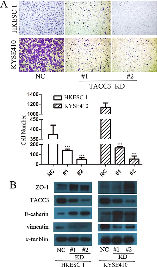
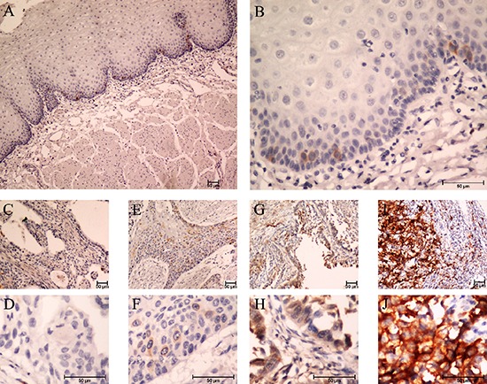
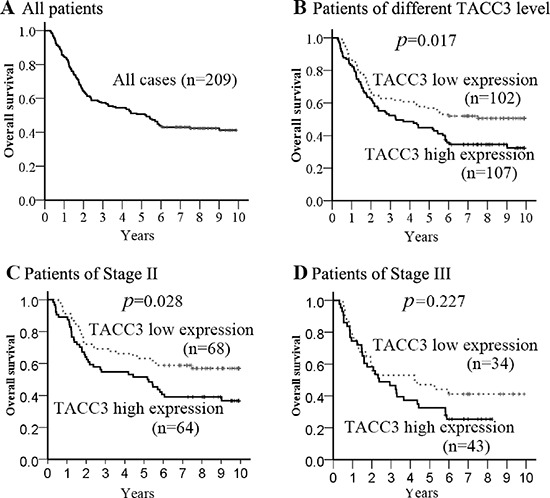
To analyze the expression of the transforming acidic coiled-coil protein 3 (TACC3) in esophageal squamous cell carcinoma (ESCC) samples, and to identify whether TACC3 can serve as a biomarker for the diagnosis and prognosis of ESCC, qPCR, western blotting and immunohistochemistry staining (IHC) were utilized to detect the expression of TACC3. Furthermore, cell growth, colony formation, migration ability and the epithelial-mesenchymal transition markers of ESCC cells in which TACC3 were knocked-down were measured. The mRNA and protein levels of TACC3 were higher in ESCC specimens compared to non-tumorous esophageal epithelial tissues. IHC results revealed TACC3 expression was significantly correlated to differentiation (p = 0.017) and lymphoid nodal status (p = 0.028). The patients with high-expression of TACC3 had a significantly poor prognosis compared to those of low-expression (p = 0.017), especially in the patients at stages I-II (p = 0.028). Multivariate analysis indicated that TACC3 expression was an independent prognostic factor for ESCC patients (p = 0.025). Knockdown of TACC3 inhibited the ability of cell proliferation, colony formation and migration. This study first identifies TACC3 not only as a useful biomarker for diagnose and prognosis of ESCC, but also as a potential therapeutic target for patients with ESCC.
为分析转化酸性卷曲螺旋蛋白3(TACC3)在食管鳞状细胞癌(ESCC)样本中的表达情况,并确定TACC3是否可作为ESCC诊断和预后的生物标志物,采用定量聚合酶链反应(qPCR)、蛋白质免疫印迹法和免疫组织化学染色(IHC)检测TACC3的表达。此外,还检测了TACC3基因敲低的ESCC细胞的生长、集落形成、迁移能力及上皮-间质转化标志物。与非肿瘤性食管上皮组织相比,ESCC标本中TACC3的mRNA和蛋白质水平更高。免疫组织化学结果显示,TACC3表达与分化程度(p = 0.017)和淋巴结状态(p = 0.028)显著相关。与低表达患者相比,TACC3高表达患者的预后明显较差(p = 0.017),尤其是在I-II期患者中(p = 0.028)。多因素分析表明,TACC3表达是ESCC患者的独立预后因素(p = 0.025)。敲低TACC3可抑制细胞增殖、集落形成和迁移能力。本研究首次确定TACC3不仅是ESCC诊断和预后的有用生物标志物,也是ESCC患者的潜在治疗靶点。