Thieme Karina, Oliveira-Souza Maria
Department of Physiology and Biophysics, Institute of Biomedical Sciences, University of São Paulo, São Paulo, Brazil.
PLoS One. 2015 Mar 20;10(3):e0122265. doi: 10.1371/journal.pone.0122265. eCollection 2015.
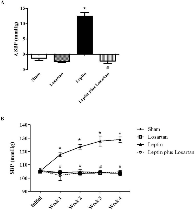
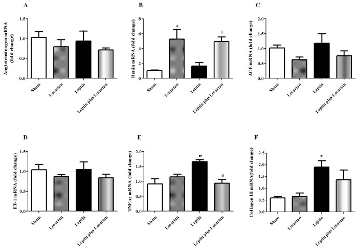
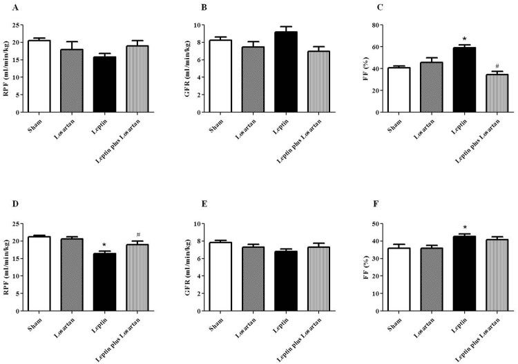
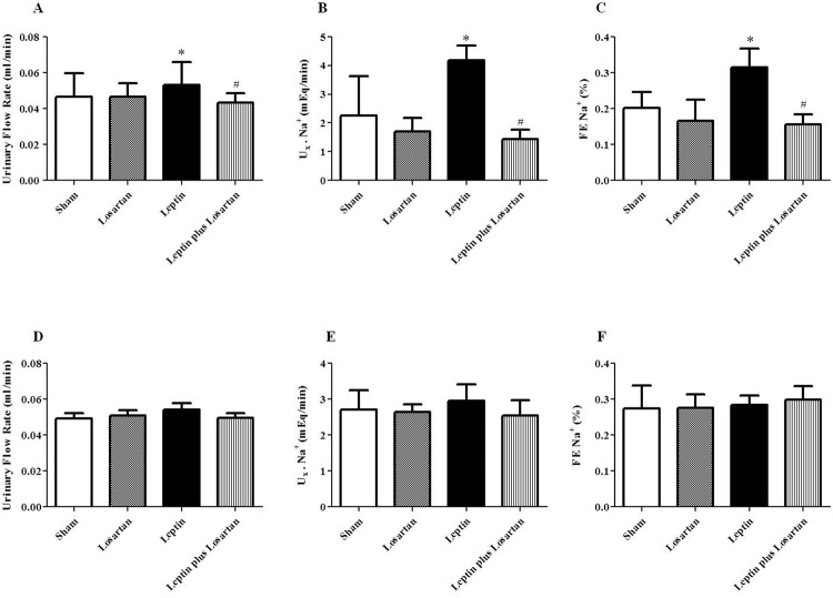
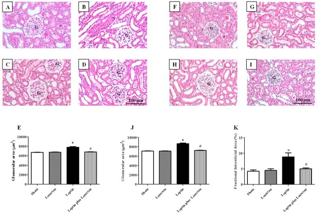
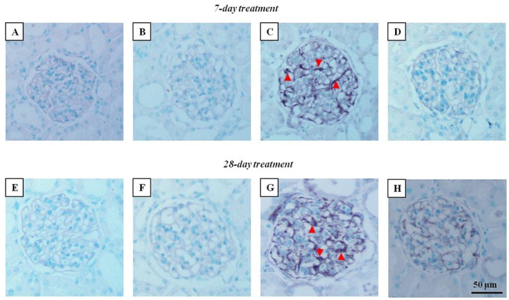
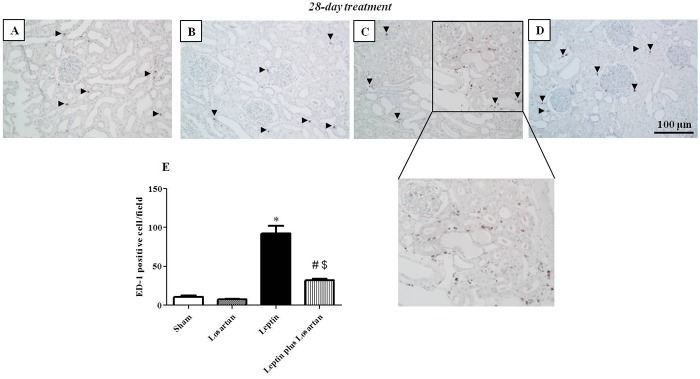
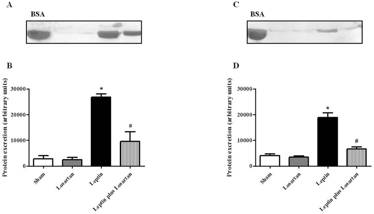
The role of hyperleptinemia in cardiovascular diseases is well known; however, in the renal tissue, the exact site of leptin's action has not been established. This study was conducted to assess the effect of leptin treatment for 7 and 28 days on renal function and morphology and the participation of angiotensin II (Ang II), through its AT1 receptor. Rats were divided into four groups: sham, losartan (10 mg/kg/day, s.c.), leptin (0.5 mg/kg/day for the 7 days group and 0.25 mg/kg/day for the 28 days group) and leptin plus losartan. Plasma leptin, Ang II and endothelin 1 (ET-1) levels were measured using an enzymatic immuno assay. The systolic blood pressure (SBP) was evaluated using the tail-cuff method. The renal plasma flow (RPF) and the glomerular filtration rate (GFR) were determined by p-aminohippuric acid and inulin clearance, respectively. Urinary Na+ and K+ levels were also analyzed. Renal morphological analyses, desmin and ED-1 immunostaining were performed. Proteinuria was analyzed by silver staining. mRNA expression of renin-angiotensin system (RAS) components, TNF-α and collagen type III was analyzed by quantitative PCR. Our results showed that leptin treatment increased Ang II plasma levels and progressively increased the SBP, achieving a pre-hypertension state. Rats treated with leptin 7 days showed a normal RPF and GFR, but increased filtration fraction (FF) and natriuresis. However, rats treated with leptin for 28 showed a decrease in the RPF, an increase in the FF and no changes in the GFR or tubular function. Leptin treatment-induced renal injury was demonstrated by: glomerular hypertrophy, increased desmin staining, macrophage infiltration in the renal tissue, TNF-α and collagen type III mRNA expression and proteinuria. In conclusion, our study demonstrated the progressive renal morphological changes in experimental hyperleptinemia and the interaction between leptin and the RAS on these effects.
高瘦素血症在心血管疾病中的作用已广为人知;然而,在肾组织中,瘦素的确切作用位点尚未明确。本研究旨在评估瘦素治疗7天和28天对肾功能和形态的影响,以及血管紧张素II(Ang II)通过其AT1受体所起的作用。将大鼠分为四组:假手术组、氯沙坦组(10毫克/千克/天,皮下注射)、瘦素组(7天组为0.5毫克/千克/天,28天组为0.25毫克/千克/天)以及瘦素加氯沙坦组。采用酶免疫分析法测定血浆瘦素、Ang II和内皮素1(ET-1)水平。使用尾套法评估收缩压(SBP)。分别通过对氨基马尿酸和菊粉清除率测定肾血浆流量(RPF)和肾小球滤过率(GFR)。还分析了尿钠和钾水平。进行了肾脏形态学分析、结蛋白和ED-1免疫染色。通过银染分析蛋白尿。采用定量PCR分析肾素-血管紧张素系统(RAS)成分、TNF-α和III型胶原的mRNA表达。我们的结果显示,瘦素治疗可提高血浆Ang II水平,并使SBP逐渐升高,达到高血压前期状态。接受7天瘦素治疗的大鼠RPF和GFR正常,但滤过分数(FF)和尿钠排泄增加。然而,接受28天瘦素治疗的大鼠RPF降低,FF升高,GFR或肾小管功能无变化。瘦素治疗引起的肾损伤表现为:肾小球肥大、结蛋白染色增加、肾组织巨噬细胞浸润、TNF-α和III型胶原mRNA表达以及蛋白尿。总之,我们的研究证明了实验性高瘦素血症中肾脏形态的渐进性变化,以及瘦素与RAS在这些影响上的相互作用。