Mougeolle Alexis, Poussard Sylvie, Decossas Marion, Lamaze Christophe, Lambert Olivier, Dargelos Elise
Univ Bordeaux, Chimie et Biologie des Membranes et Nanoobjets, UMR 5248, F-33600 Pessac, France; CNRS, Chimie et Biologie des Membranes et Nanoobjets, UMR 5248, F-33600 Pessac, France; Bordeaux INP, Chimie et Biologie des Membranes et Nanoobjets, UMR 5248, F-33600 Pessac, France.
Institut Curie-Centre de Recherche, Membrane Dynamics and Mechanics of Intracellular Signaling Team, INSERM U1143, CNRS UMR 3666, Paris, France.
PLoS One. 2015 Mar 23;10(3):e0122654. doi: 10.1371/journal.pone.0122654. eCollection 2015.
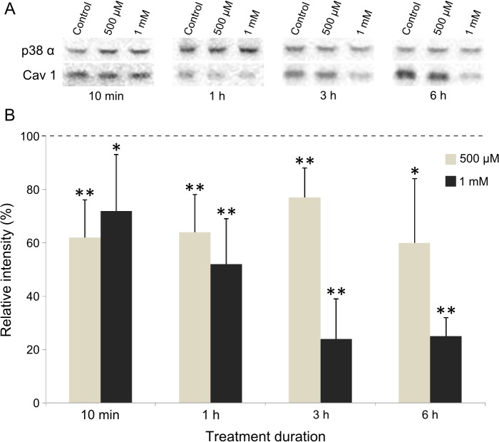
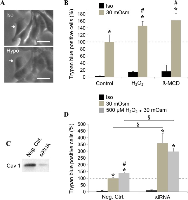
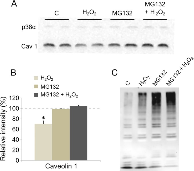
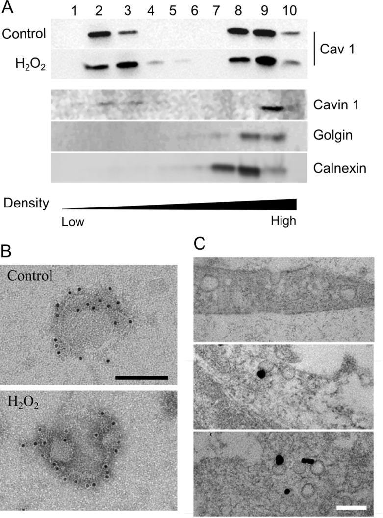
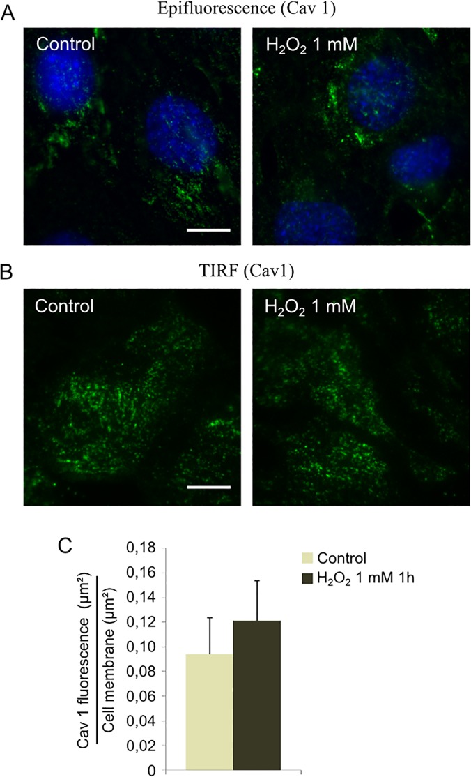
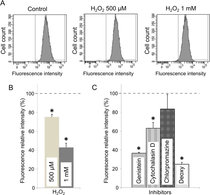
Increased level of oxidative stress, a major actor of cellular aging, impairs the regenerative capacity of skeletal muscle and leads to the reduction in the number and size of muscle fibers causing sarcopenia. Caveolin 1 is the major component of caveolae, small membrane invaginations involved in signaling and endocytic trafficking. Their role has recently expanded to mechanosensing and to the regulation of oxidative stress-induced pathways. Here, we increased the amount of reactive oxidative species in myoblasts by addition of hydrogen peroxide (H2O2) at non-toxic concentrations. The expression level of caveolin 1 was significantly decreased as early as 10 min after 500 μM H2O2 treatment. This reduction was not observed in the presence of a proteasome inhibitor, suggesting that caveolin 1 was rapidly degraded by the proteasome. In spite of caveolin 1 decrease, caveolae were still able to assemble at the plasma membrane. Their functions however were significantly perturbed by oxidative stress. Endocytosis of a ceramide analog monitored by flow cytometry was significantly diminished after H2O2 treatment, indicating that oxidative stress impaired its selective internalization via caveolae. The contribution of caveolae to the plasma membrane reservoir has been monitored after osmotic cell swelling. H2O2 treatment increased membrane fragility revealing that treated cells were more sensitive to an acute mechanical stress. Altogether, our results indicate that H2O2 decreased caveolin 1 expression and impaired caveolae functions. These data give new insights on age-related deficiencies in skeletal muscle.
氧化应激水平升高是细胞衰老的主要因素,会损害骨骼肌的再生能力,并导致肌纤维数量和大小减少,从而引起肌肉减少症。小窝蛋白1是小窝的主要成分,小窝是参与信号传导和内吞运输的小膜内陷结构。它们的作用最近已扩展到机械传感以及氧化应激诱导途径的调节。在这里,我们通过添加无毒浓度的过氧化氢(H2O2)来增加成肌细胞中活性氧的量。在500μM H2O2处理后仅10分钟,小窝蛋白1的表达水平就显著降低。在蛋白酶体抑制剂存在的情况下未观察到这种降低,这表明小窝蛋白1被蛋白酶体迅速降解。尽管小窝蛋白1减少,但小窝仍能够在质膜上组装。然而,它们的功能受到氧化应激的显著干扰。通过流式细胞术监测的神经酰胺类似物的内吞作用在H2O2处理后显著减弱,表明氧化应激损害了其通过小窝的选择性内化。在渗透性细胞肿胀后监测了小窝对质膜储备的贡献。H2O2处理增加了膜的脆性,表明处理后的细胞对急性机械应激更敏感。总之,我们的结果表明H2O2降低了小窝蛋白1的表达并损害了小窝的功能。这些数据为骨骼肌中与年龄相关的缺陷提供了新的见解。